
















©The Author(s) 2026.
World J Gastrointest Oncol. Jan 15, 2026; 18(1): 114648
Published online Jan 15, 2026. doi: 10.4251/wjgo.v18.i1.114648
Published online Jan 15, 2026. doi: 10.4251/wjgo.v18.i1.114648
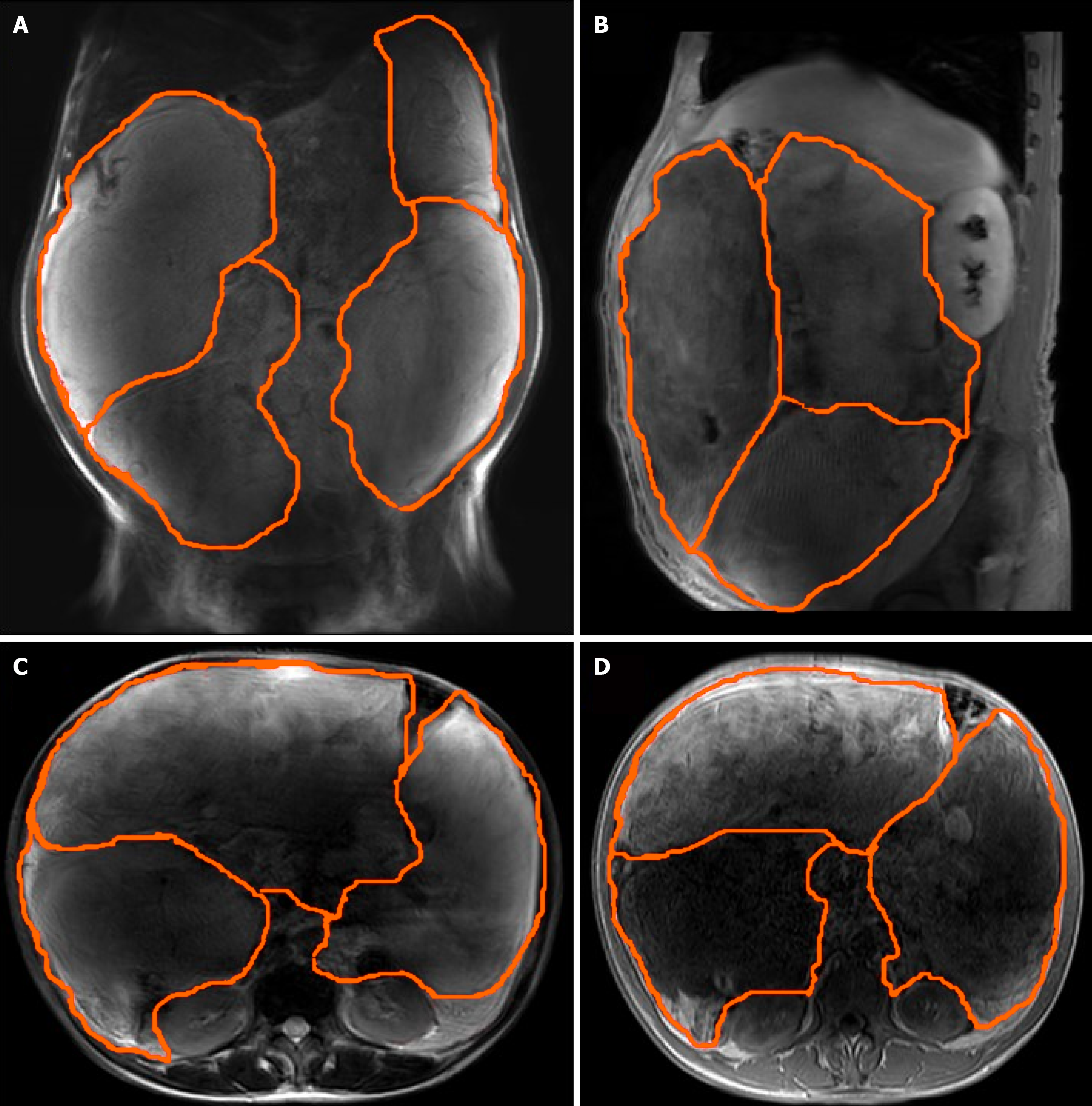
Figure 2 Abdominal and pelvic magnetic resonance imaging scan indicated that multiple round masses were found in the abdominal cavity with an envelope, and occupy the entire abdominal cavity, which originated from the small bowel mesentery.
Tremendous masses, well circumscribed, lobulated, heterogeneous mass, compartmentalized, with a maximum size of 25 cm × 23 cm × 15 cm (mark with orange lines). A: Coronal position; B: Sagittal position; C: Transverse position T2 weighted image; D: Transverse position T1 weighted image.
- Citation: Tian Y, Liu GQ, Li CF, Tian QM, Qiao S. Colossal well-differentiated liposarcoma of the small bowel mesentery: A case report. World J Gastrointest Oncol 2026; 18(1): 114648
- URL: https://www.wjgnet.com/1948-5204/full/v18/i1/114648.htm
- DOI: https://dx.doi.org/10.4251/wjgo.v18.i1.114648