
















©The Author(s) 2026.
World J Gastrointest Oncol. Jan 15, 2026; 18(1): 114339
Published online Jan 15, 2026. doi: 10.4251/wjgo.v18.i1.114339
Published online Jan 15, 2026. doi: 10.4251/wjgo.v18.i1.114339
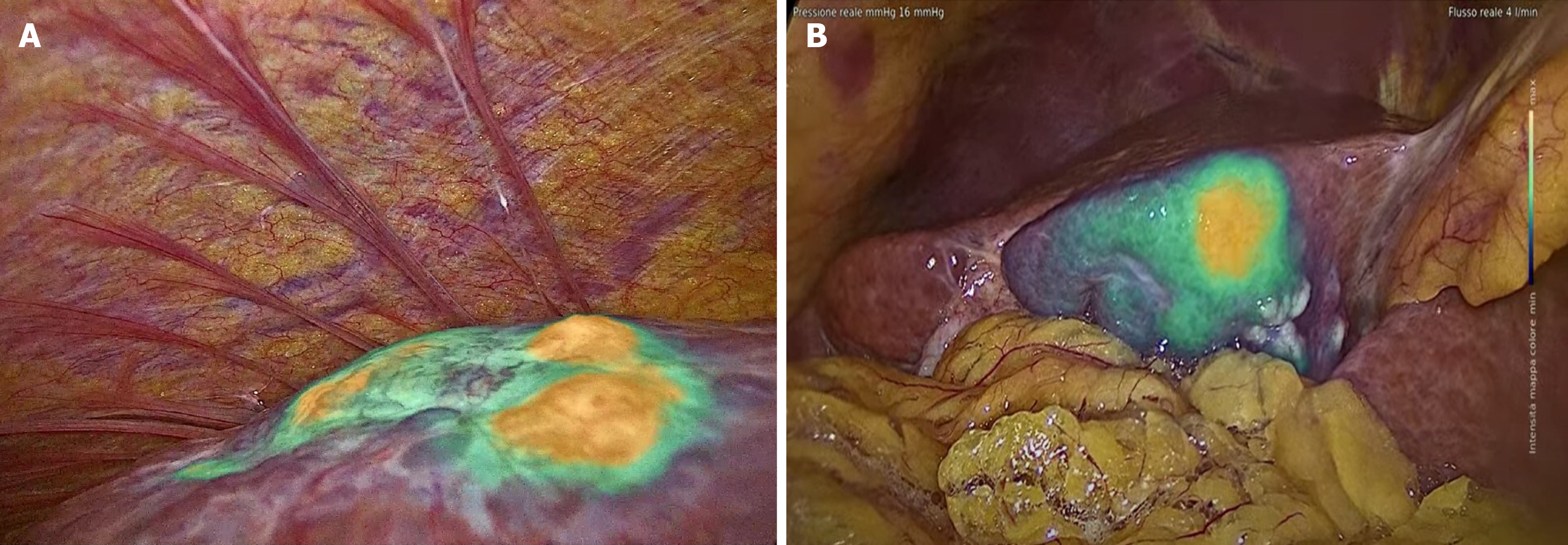
Figure 2 Indocyanine green fluorescence patterns and microinvasive components.
A: Hepatocellular carcinoma with vascular micro-invasion, indocyanine green (ICG)-window view showed more nodules with intense ICG fluoresce; B: Hepatocellular carcinoma with microinvasive components due to periductal infiltrating growth, which showed a homogeneous and intense area of neoplastic tissue with partial fluorescence around the peritumoral area (intensity maps of IMAGE1 S™ Rubina®, Karl Storz Endoscope).
- Citation: Piccolo G, Barabino M, Benuzzi L, Formisano G, Bianchi PP. Clinical applications of indocyanine green fluorescence for the treatment of hepatocellular carcinoma. World J Gastrointest Oncol 2026; 18(1): 114339
- URL: https://www.wjgnet.com/1948-5204/full/v18/i1/114339.htm
- DOI: https://dx.doi.org/10.4251/wjgo.v18.i1.114339