
















©The Author(s) 2026.
World J Gastrointest Oncol. Jan 15, 2026; 18(1): 111144
Published online Jan 15, 2026. doi: 10.4251/wjgo.v18.i1.111144
Published online Jan 15, 2026. doi: 10.4251/wjgo.v18.i1.111144
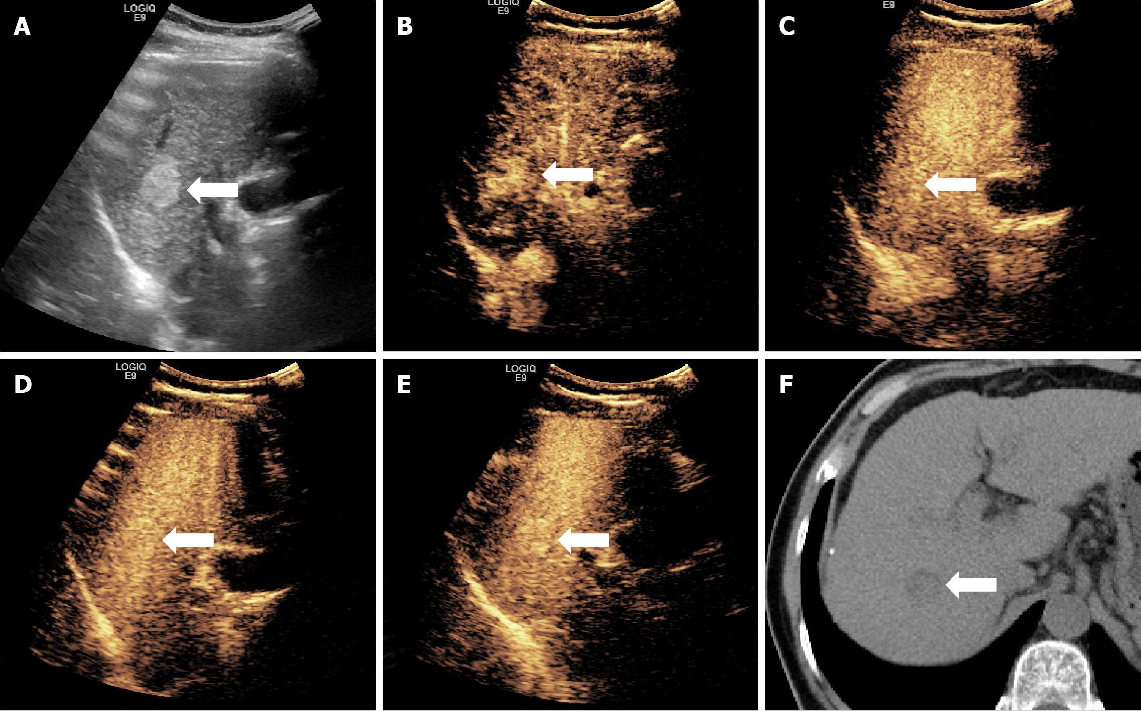
Figure 5 Ultrasound and computed tomography figures of a case of hepatocellular carcinoma showing hyper-enhancement in Kupffer phase.
A typical figure presents the case of a 57-year-old male patient with chronic hepatitis B, in whom a liver mass was detected during a physical examination. After surgical resection, pathology of the lesion revealed well-differentiated hepatocellular carcinoma without vascular invasion, and a Ki-67 LI of about 3%. A: An ultrasound image showing a mass approximately 2.1 cm in diameter located in the right lobe; B-E: Sonazoid-contrast-enhanced ultrasonography images show rapid enhancement of the mass in arterial phase, and washout does not occur in the following three phases; F: Computed tomography plain image revealing the mass.
- Citation: Liu RB, Xin JY, Huang Z, Li KY. Sonazoid-contrast-enhanced ultrasound for the histological diagnosis of hepatocellular carcinoma. World J Gastrointest Oncol 2026; 18(1): 111144
- URL: https://www.wjgnet.com/1948-5204/full/v18/i1/111144.htm
- DOI: https://dx.doi.org/10.4251/wjgo.v18.i1.111144