
















©The Author(s) 2025.
World J Gastrointest Oncol. Sep 15, 2025; 17(9): 109055
Published online Sep 15, 2025. doi: 10.4251/wjgo.v17.i9.109055
Published online Sep 15, 2025. doi: 10.4251/wjgo.v17.i9.109055
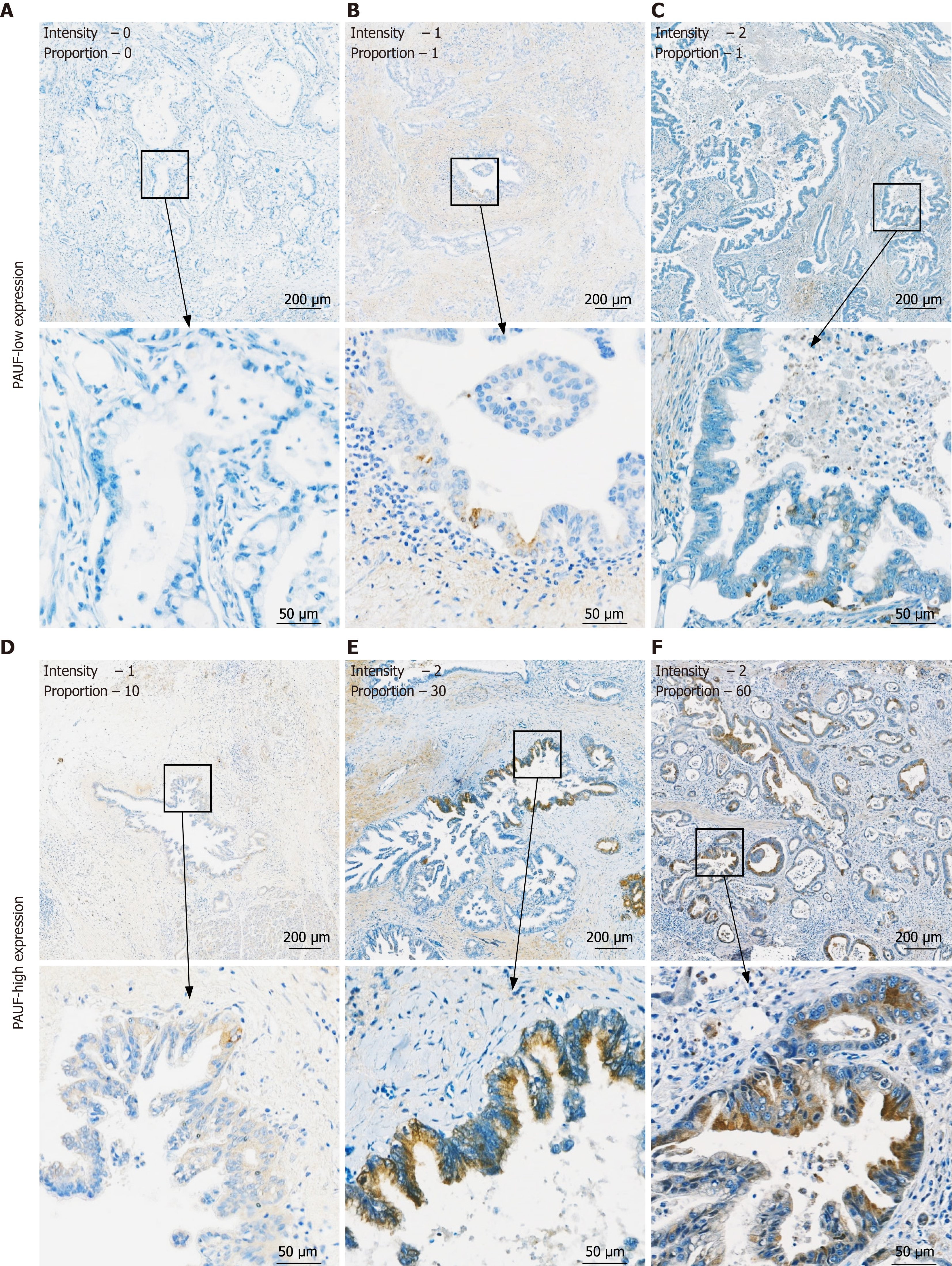
Figure 2 Immunohistochemistry staining results of pancreatic adenocarcinoma upregulated factor in pancreatic cancer patient tissues.
A-F: Representative immunohistochemistry images from six pancreatic cancer tissue samples demonstrating varying levels of pancreatic adenocarcinoma upregulated factor (PAUF) expression, assessed by both staining intensity and the proportion of PAUF-positive tumor cells. Samples classified as PAUF-low exhibit staining intensity of 0 or 1+ with ≤ 4% of tumor cells stained, while PAUF-high cases show staining intensity of 1+ or 2+ with > 5% of tumor cells stained. PAUF: Pancreatic adenocarcinoma upregulated factor.
- Citation: Kim JH, Na HY, Jung K, Jang D, Youn Y, Kim DH, Han HD, Hwang JH. Quantitative immunohistochemistry analysis of pancreatic adenocarcinoma upregulated factor expression in pancreatic cancer and its prognostic significance. World J Gastrointest Oncol 2025; 17(9): 109055
- URL: https://www.wjgnet.com/1948-5204/full/v17/i9/109055.htm
- DOI: https://dx.doi.org/10.4251/wjgo.v17.i9.109055