Copyright
©The Author(s) 2025.
World J Gastrointest Oncol. Jul 15, 2025; 17(7): 108650
Published online Jul 15, 2025. doi: 10.4251/wjgo.v17.i7.108650
Published online Jul 15, 2025. doi: 10.4251/wjgo.v17.i7.108650
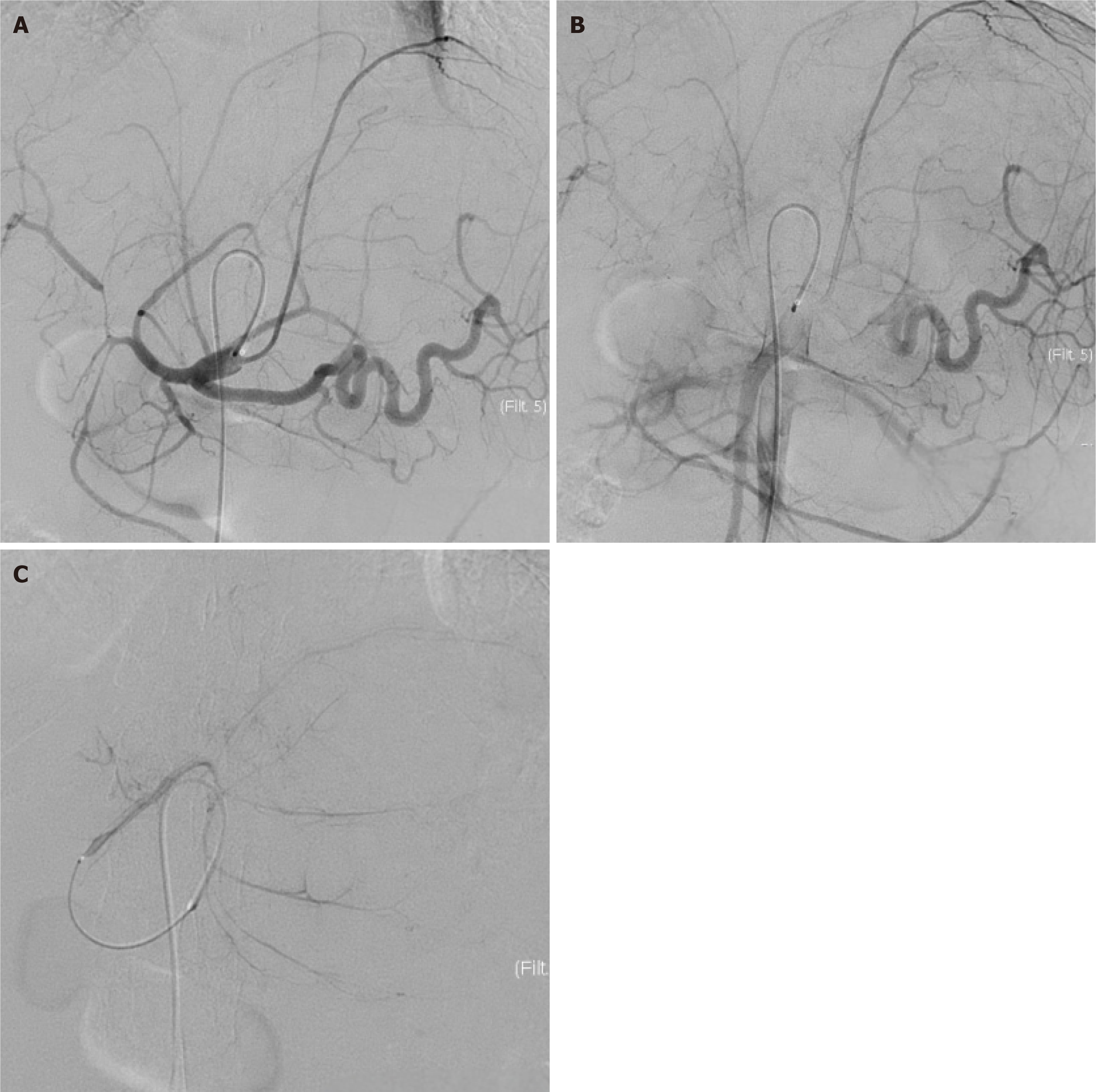
Figure 7 Pre-chemotherapy hepatic arteriography.
A and B: The left hepatic artery is the dominant feeding artery of the tumor, demonstrating marked luminal dilation, serpentine tortuosity, and proliferative branching encircling the tumor in the early and middle arterial phases; C: Prominent tumor staining is observed in the late arterial phase during microcatheter angiography.
- Citation: Xie JP, Tang YJ, Fan YW, Huang YZ, Deng M, Zhang TZ, Li Y, Deng G, Tang D. Pathological complete response in advanced intrahepatic cholangiocarcinoma was achieved through tri-modal therapy: A case report and review of literature. World J Gastrointest Oncol 2025; 17(7): 108650
- URL: https://www.wjgnet.com/1948-5204/full/v17/i7/108650.htm
- DOI: https://dx.doi.org/10.4251/wjgo.v17.i7.108650


