Copyright
©The Author(s) 2025.
World J Gastrointest Oncol. Jul 15, 2025; 17(7): 108650
Published online Jul 15, 2025. doi: 10.4251/wjgo.v17.i7.108650
Published online Jul 15, 2025. doi: 10.4251/wjgo.v17.i7.108650
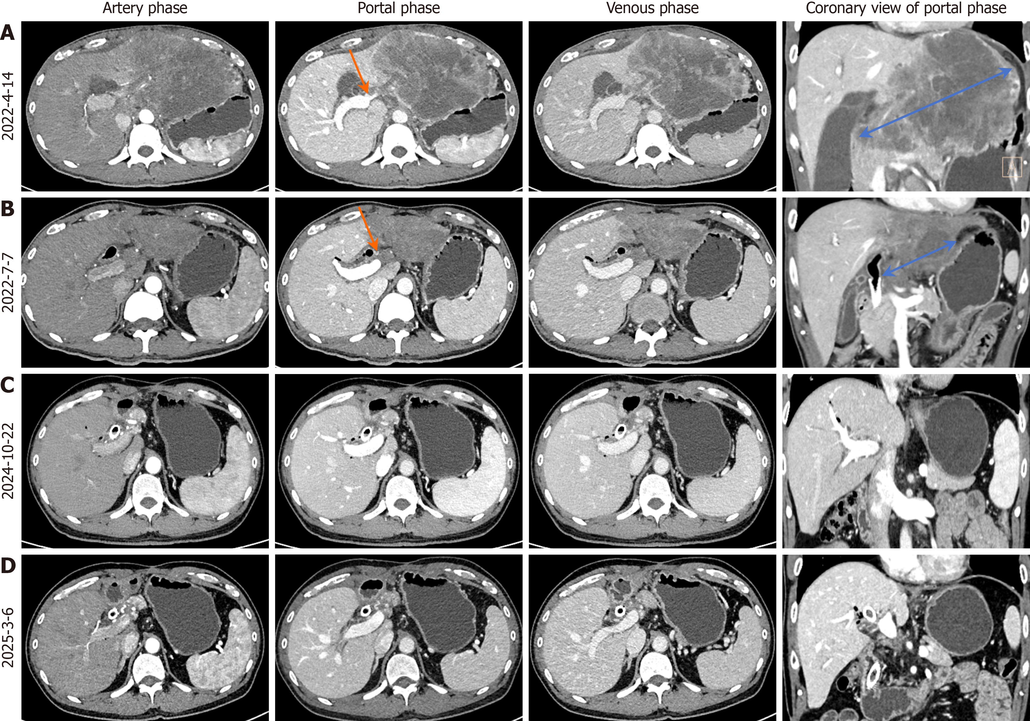
Figure 1 Serial contrast-enhanced computed tomography imaging during the treatment course.
A: Pre-chemotherapy baseline computed tomography (CT) showing a 13-cm hypodense mass (blue arrow) with heterogeneous enhancement in the left hepatic lobe, exhibiting mild to moderate peripheral enhancement and left portal branch encasement (orange arrow); B: Follow-up CT after 3 cycles of combined therapy demonstrating a significant reduction in tumor diameter to 6.5 cm (blue arrow), with attenuated enhancement compared to baseline and alleviated left portal branch involvement (orange arrow); C: Post-resection CT at 24-month follow-up revealing no evidence of tumor recurrence; D: Post-resection CT at 29-month follow-up confirming the absence of tumor recurrence.
- Citation: Xie JP, Tang YJ, Fan YW, Huang YZ, Deng M, Zhang TZ, Li Y, Deng G, Tang D. Pathological complete response in advanced intrahepatic cholangiocarcinoma was achieved through tri-modal therapy: A case report and review of literature. World J Gastrointest Oncol 2025; 17(7): 108650
- URL: https://www.wjgnet.com/1948-5204/full/v17/i7/108650.htm
- DOI: https://dx.doi.org/10.4251/wjgo.v17.i7.108650


