Copyright
©The Author(s) 2025.
World J Gastrointest Oncol. Jul 15, 2025; 17(7): 108258
Published online Jul 15, 2025. doi: 10.4251/wjgo.v17.i7.108258
Published online Jul 15, 2025. doi: 10.4251/wjgo.v17.i7.108258
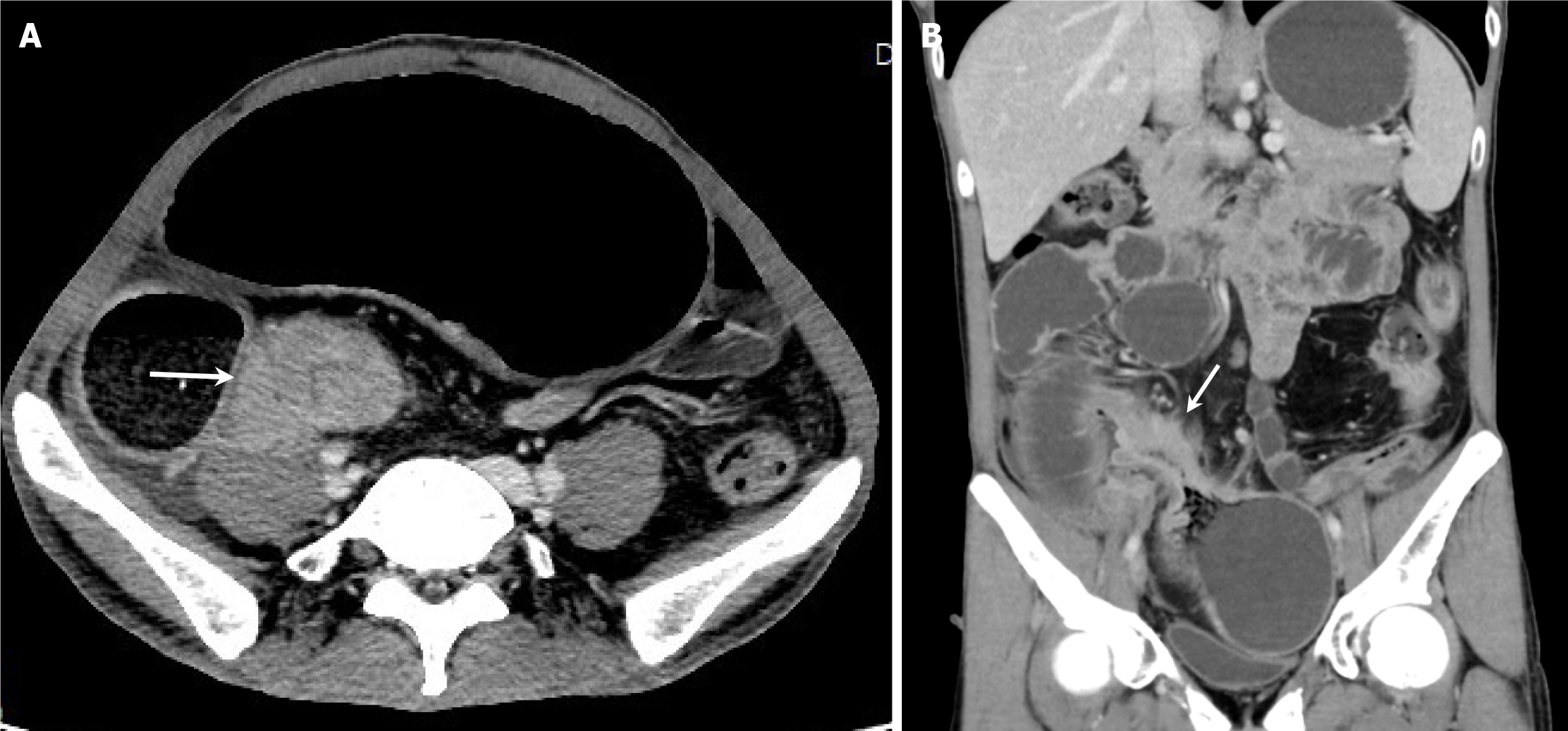
Figure 2 Computed tomography enterography images of the patient.
A and B: Computed tomography enterography demonstrates segmental bowel wall thickening, local narrowing of the intestinal lumen, dilation and fluid accumulation in the small intestine, clear surrounding fat planes, and slight increase in the number of vessels on the mesenteric side.
- Citation: Zhong MY, Jian GL, Ye JY, Chen KX, Huang WJ. Ultrasound diagnosis of small bowel adenocarcinoma in Crohn’s disease: A case report and review of literature. World J Gastrointest Oncol 2025; 17(7): 108258
- URL: https://www.wjgnet.com/1948-5204/full/v17/i7/108258.htm
- DOI: https://dx.doi.org/10.4251/wjgo.v17.i7.108258


