Copyright
©The Author(s) 2025.
World J Gastrointest Oncol. Jul 15, 2025; 17(7): 108258
Published online Jul 15, 2025. doi: 10.4251/wjgo.v17.i7.108258
Published online Jul 15, 2025. doi: 10.4251/wjgo.v17.i7.108258
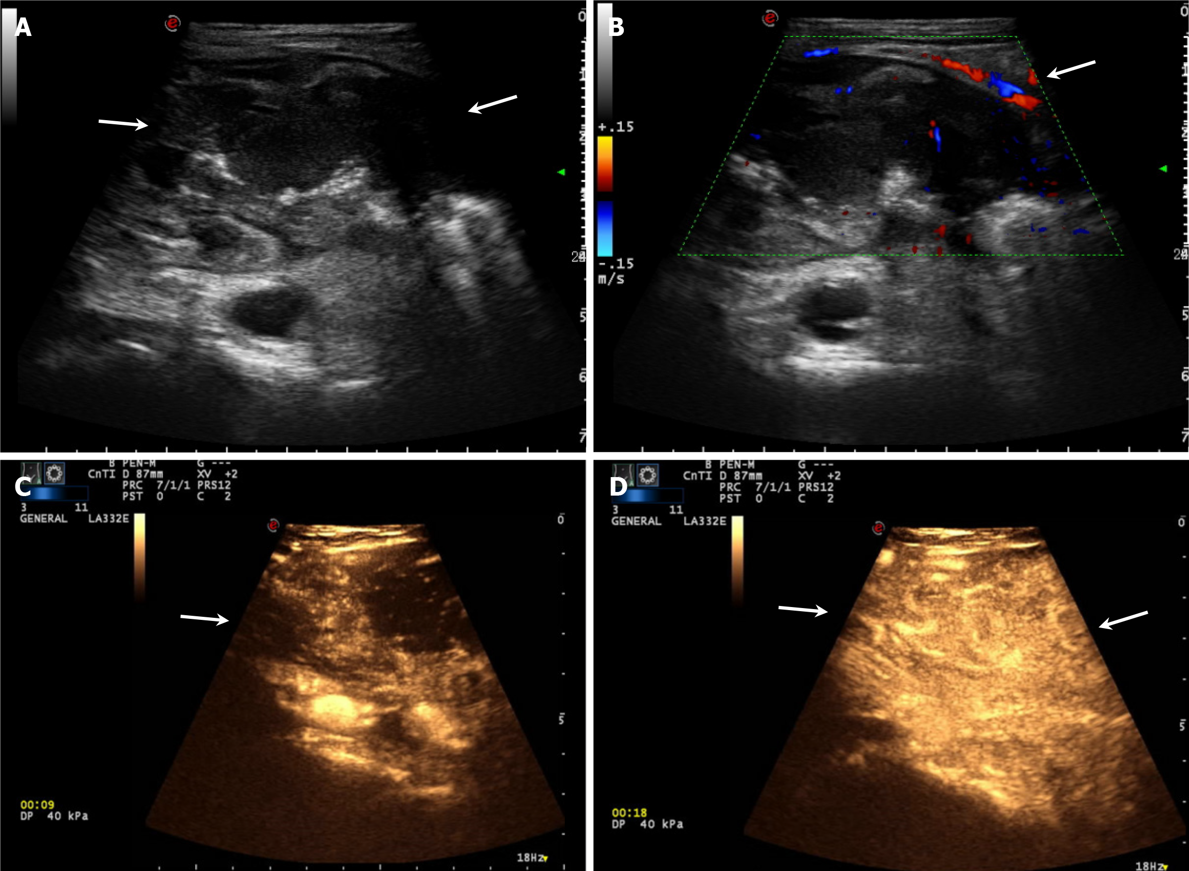
Figure 1 Ultrasound images of the patient.
A: Intestinal ultrasonography demonstrated uneven, eccentric wall thickening of the ileocecal junction, disappearance of the layered structure, and the pseudo-kidney sign (arrows); B: Color Doppler Flow Imaging revealed linear blood flow (arrows); C and D: After a bolus injection of 2.0 mL of Sonovue contrast agent into the left antecubital vein of the patient, contrast-enhanced ultrasonography demonstrated disappearance of the bowel wall stratification, heterogeneous high enhancement, and rapid washout (arrows).
- Citation: Zhong MY, Jian GL, Ye JY, Chen KX, Huang WJ. Ultrasound diagnosis of small bowel adenocarcinoma in Crohn’s disease: A case report and review of literature. World J Gastrointest Oncol 2025; 17(7): 108258
- URL: https://www.wjgnet.com/1948-5204/full/v17/i7/108258.htm
- DOI: https://dx.doi.org/10.4251/wjgo.v17.i7.108258


