Copyright
©The Author(s) 2025.
World J Gastrointest Oncol. Jul 15, 2025; 17(7): 108162
Published online Jul 15, 2025. doi: 10.4251/wjgo.v17.i7.108162
Published online Jul 15, 2025. doi: 10.4251/wjgo.v17.i7.108162
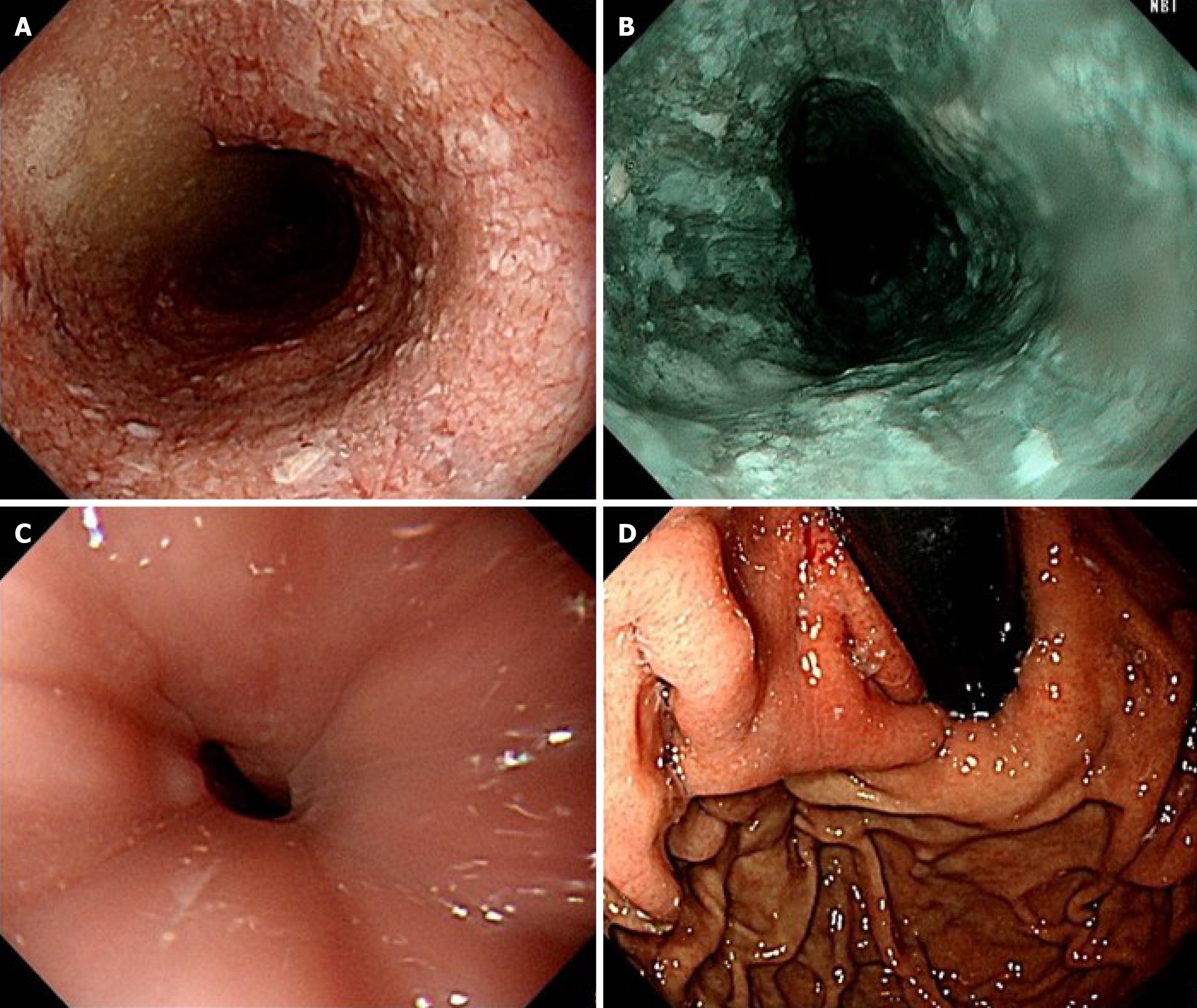
Figure 1 Esophagogastroduodenoscopy.
A: Food and fluid accumulation in the dilated esophagus; B: No abnormal mucosa under narrow-band imaging; C: Tight esophagogastric junction; D: The retroflexion examination revealed no tumors in the gastric cardia.
- Citation: He YS, Lee CY, Shieh TY. Pseudoachalasia as first manifestation of a diffusely infiltrative esophageal squamous cell carcinoma: A case report. World J Gastrointest Oncol 2025; 17(7): 108162
- URL: https://www.wjgnet.com/1948-5204/full/v17/i7/108162.htm
- DOI: https://dx.doi.org/10.4251/wjgo.v17.i7.108162


