Copyright
©The Author(s) 2025.
World J Gastrointest Oncol. Jun 15, 2025; 17(6): 106846
Published online Jun 15, 2025. doi: 10.4251/wjgo.v17.i6.106846
Published online Jun 15, 2025. doi: 10.4251/wjgo.v17.i6.106846
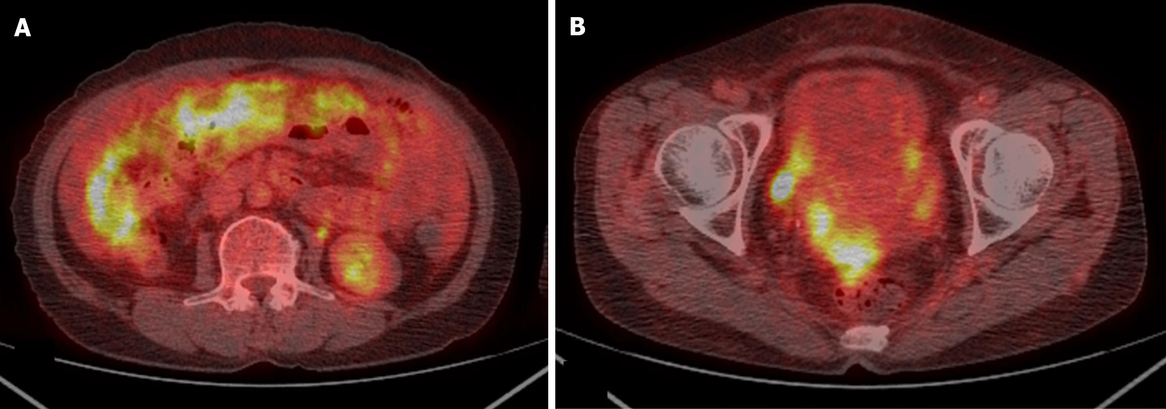
Figure 3 Positron emission tomography-computed tomography of the patient’s abdomen and pelvis.
A: Extensive hypermetabolic activity throughout the mesentery and omentum, suggesting diffuse peritoneal metastasis; B: Increased metabolic uptake in the pelvic region, indicating likely peritoneal seeding in the pelvic cavity.
- Citation: Suh JW, Jo JW, Park DG. Long-term survival of a patient with colorectal cancer with peritoneal carcinomatosis and low completeness of cytoreduction score: A case report. World J Gastrointest Oncol 2025; 17(6): 106846
- URL: https://www.wjgnet.com/1948-5204/full/v17/i6/106846.htm
- DOI: https://dx.doi.org/10.4251/wjgo.v17.i6.106846


