Copyright
©The Author(s) 2025.
World J Gastrointest Oncol. Apr 15, 2025; 17(4): 102085
Published online Apr 15, 2025. doi: 10.4251/wjgo.v17.i4.102085
Published online Apr 15, 2025. doi: 10.4251/wjgo.v17.i4.102085
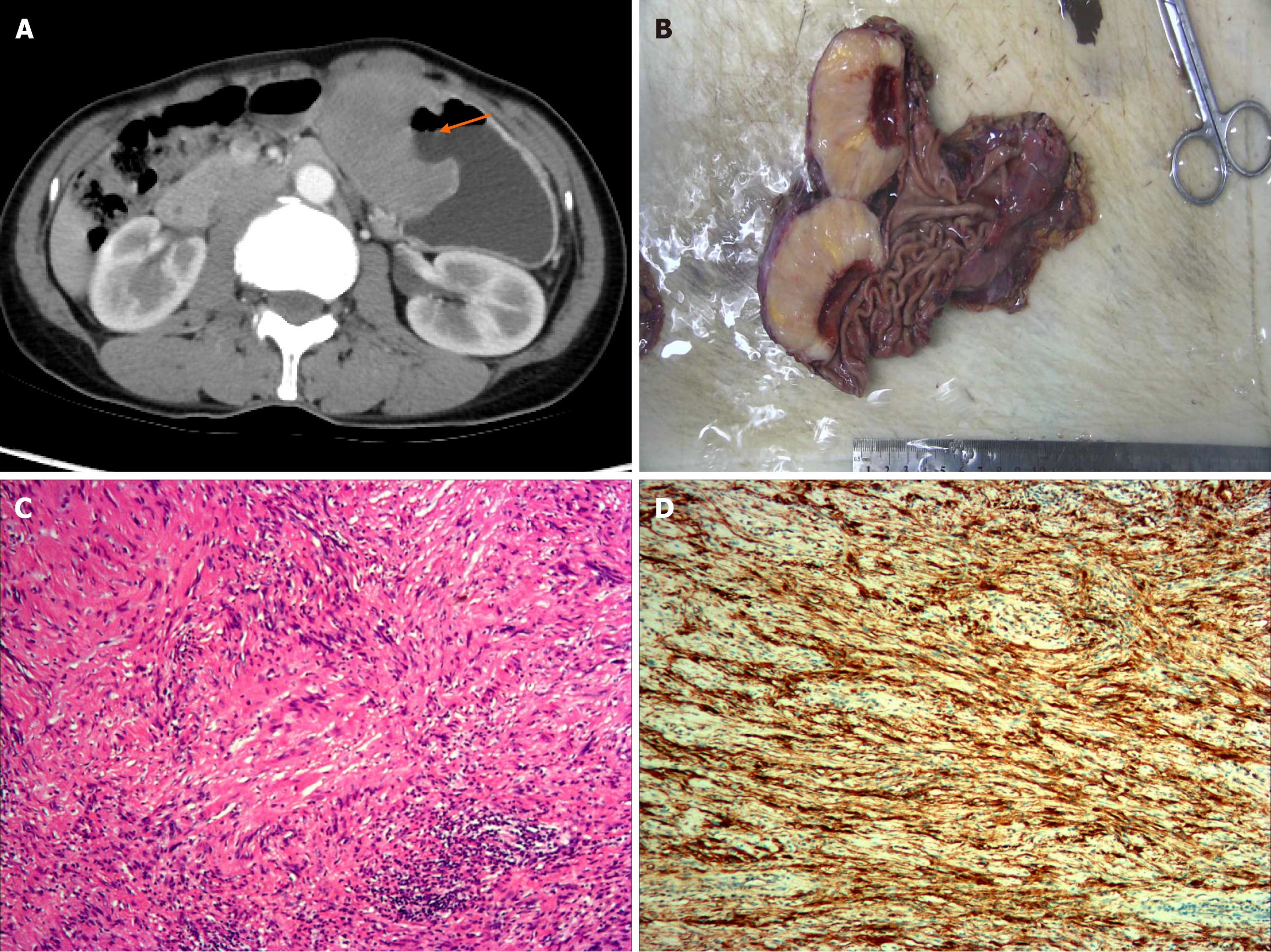
Figure 2 Gastric schwannoma in a 56-year-old woman presenting with epigastric discomfort and black stool.
A: Arterial phase-enhanced computed tomography (CT) shows an endoluminal tumor with deep ulceration (arrow); B: Gross examination revealed that the tumor’s cut surface was yellowish, with an ulcer as observed in the CT image; C: Tumor is primarily composed of spindle-shaped cells with a characteristic peripheral lymphoid cuff (hematoxylin and eosin stain; original magnification, × 100); D: The tumor is strongly positive for S-100 protein.
- Citation: Mo YK, Chen XP, Hong LL, Hu YR, Lin DY, Xie LC, Dai ZZ. Gastric schwannoma: Computed tomography and perigastric lymph node characteristics. World J Gastrointest Oncol 2025; 17(4): 102085
- URL: https://www.wjgnet.com/1948-5204/full/v17/i4/102085.htm
- DOI: https://dx.doi.org/10.4251/wjgo.v17.i4.102085


