Copyright
©The Author(s) 2025.
World J Gastrointest Oncol. Apr 15, 2025; 17(4): 102085
Published online Apr 15, 2025. doi: 10.4251/wjgo.v17.i4.102085
Published online Apr 15, 2025. doi: 10.4251/wjgo.v17.i4.102085
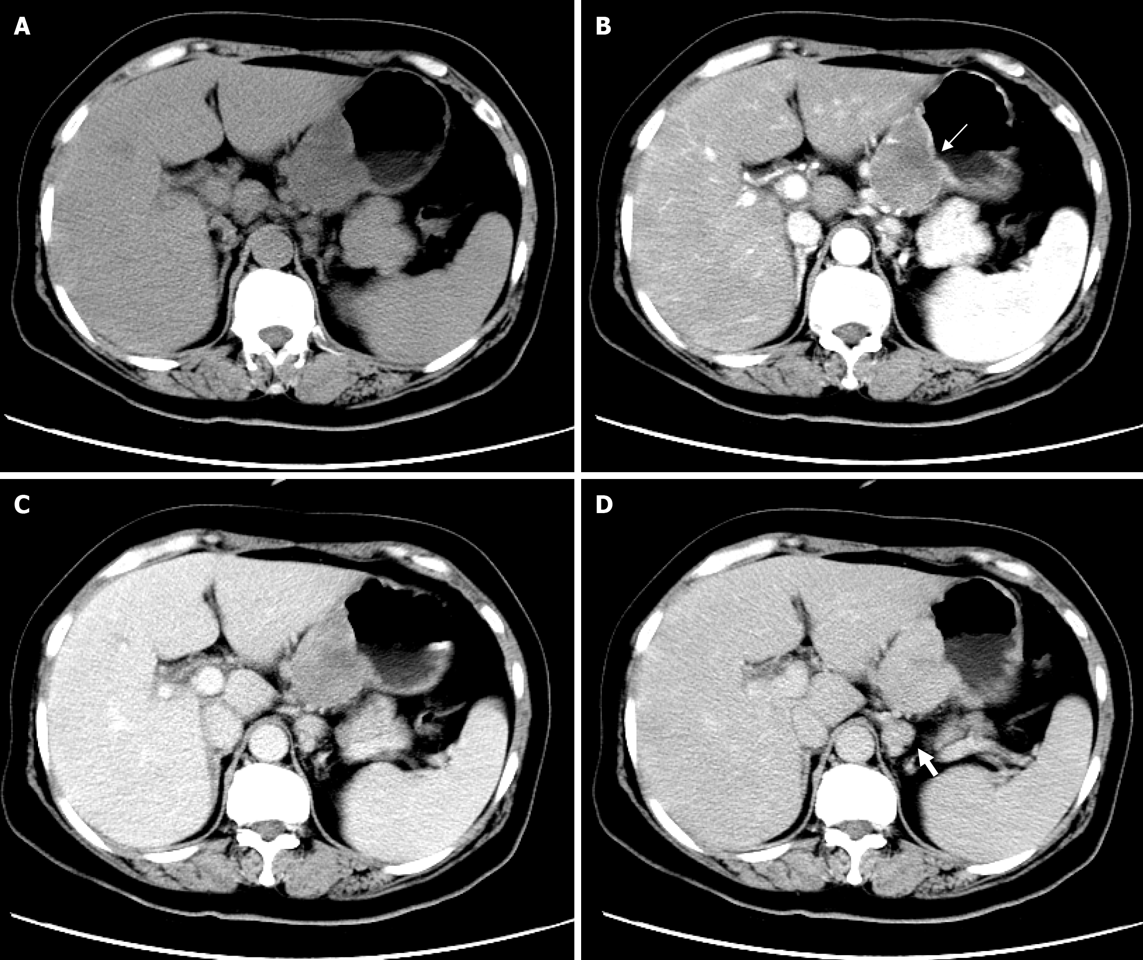
Figure 1 Gastric schwannoma in the lesser curvature of the gastric body in a 52-year-old woman.
A: Plain axial computed tomography (CT) scan shows an oval, well-defined, exophytic, and homogeneous mass, with a density of 27 Hounsfield units (HU) lower than that of the erector spinae (54 HU); B-D: Axial contrast-enhanced CT scans in the arterial, portal venous, and delayed phases show progressive homogeneous enhancement. The CT values in the three phases are 53, 66, and 70 HU, respectively. The arterial phase shows enhanced mucosal clarity, suggesting a submucosal mass. Localized mucosal disruption indicates a shallow ulcer (thin arrow). Additionally, homogeneously enhanced perigastric lymph nodes (thick arrow) are detected adjacent to the mass.
- Citation: Mo YK, Chen XP, Hong LL, Hu YR, Lin DY, Xie LC, Dai ZZ. Gastric schwannoma: Computed tomography and perigastric lymph node characteristics. World J Gastrointest Oncol 2025; 17(4): 102085
- URL: https://www.wjgnet.com/1948-5204/full/v17/i4/102085.htm
- DOI: https://dx.doi.org/10.4251/wjgo.v17.i4.102085


