
















©The Author(s) 2025.
World J Gastrointest Oncol. Apr 15, 2025; 17(4): 101123
Published online Apr 15, 2025. doi: 10.4251/wjgo.v17.i4.101123
Published online Apr 15, 2025. doi: 10.4251/wjgo.v17.i4.101123
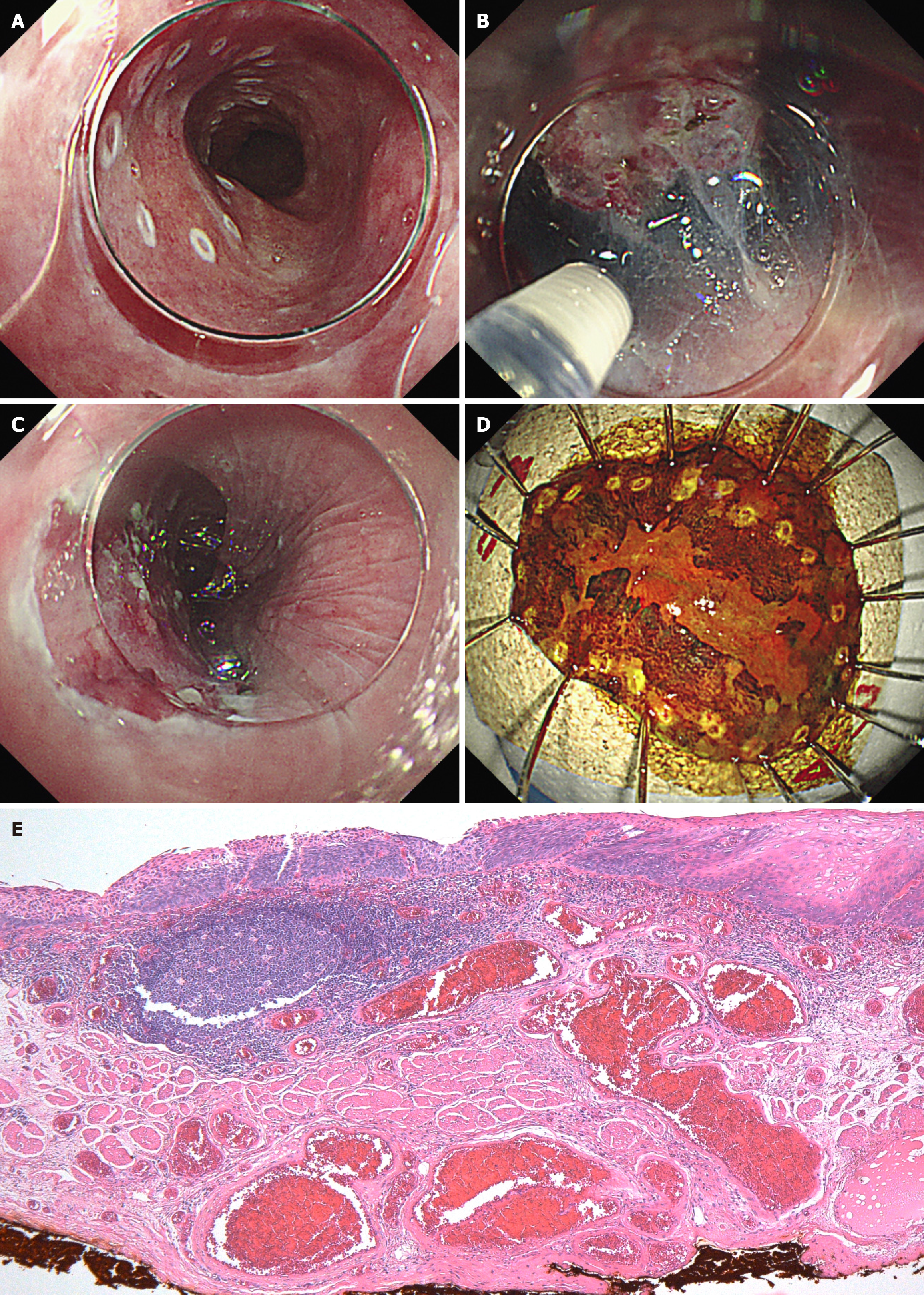
Figure 5 Endoscopic submucosal dissection.
A: Marking was performed around the lesion; B: No fibrosis was observed in the submucosal layer during endoscopic submucosal dissection; C: The lesion was completely resected without significant complications; D: Lugol's iodine staining of the lesion revealed to be removed en bloc; E: Pathological analysis revealed moderately differentiated squamous cell carcinoma and the invasion depth of the cancer was limited to lamina propria mucosa with negative vertical and horizontal margins, without lymphovascular invasion.
- Citation: Tachibana S, Moriichi K, Takahashi K, Sato M, Kobayashi Y, Sugiyama Y, Sasaki T, Sakatani A, Ando K, Ueno N, Kashima S, Tanabe H, Fujiya M. Curative endoscopic submucosal dissection for esophageal squamous cell carcinoma after chemoradiotherapy for pharyngeal cancer: A case report. World J Gastrointest Oncol 2025; 17(4): 101123
- URL: https://www.wjgnet.com/1948-5204/full/v17/i4/101123.htm
- DOI: https://dx.doi.org/10.4251/wjgo.v17.i4.101123