
















©The Author(s) 2025.
World J Gastrointest Oncol. Mar 15, 2025; 17(3): 101859
Published online Mar 15, 2025. doi: 10.4251/wjgo.v17.i3.101859
Published online Mar 15, 2025. doi: 10.4251/wjgo.v17.i3.101859
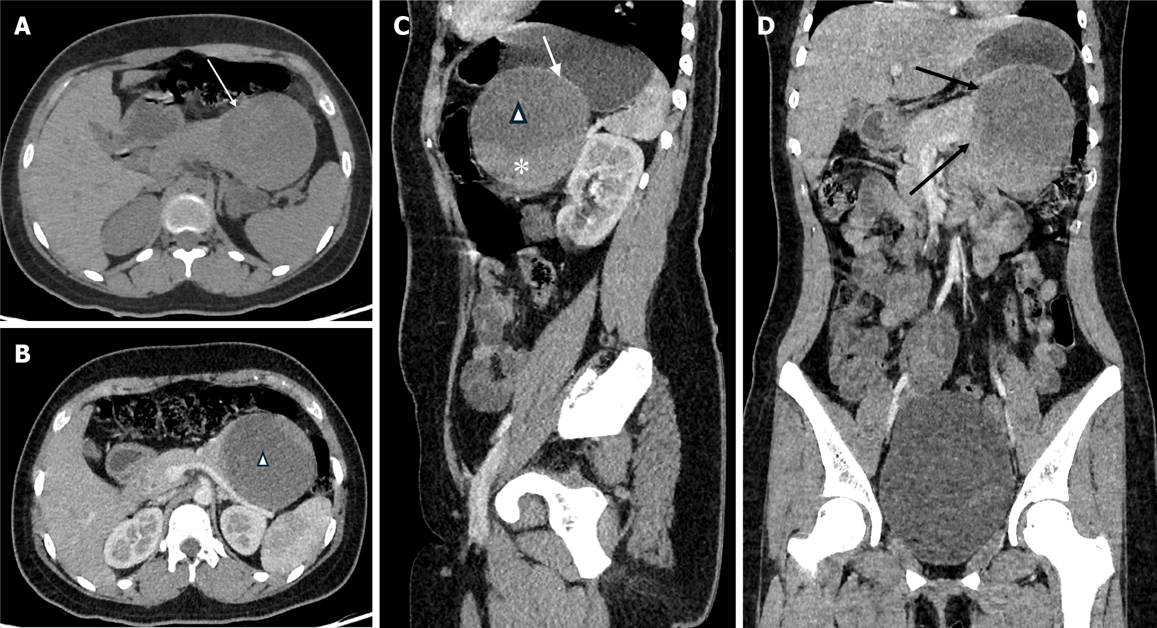
Figure 2 Computed tomography findings.
A: Non-contrast axial computed tomography image demonstrating an encapsulated, solid-cystic mass (white arrow) in the pancreatic tail; B-D: Post-contrast axial (B) with sagittal (C) view showing enhancing solid (asterisk) and non-enhancing cystic component (triangle) of the mass and coronal (D) reconstructed image showing the ‘claw sign’ formed by the mass with the pancreatic parenchyma (black arrow).
- Citation: Sapkota A, Paudel R, Pandey S, Bhatt N. Solid pseudopapillary neoplasm of the pancreas in an adolescent: A case report and review of the literature. World J Gastrointest Oncol 2025; 17(3): 101859
- URL: https://www.wjgnet.com/1948-5204/full/v17/i3/101859.htm
- DOI: https://dx.doi.org/10.4251/wjgo.v17.i3.101859