
















©The Author(s) 2025.
World J Gastrointest Oncol. Mar 15, 2025; 17(3): 100152
Published online Mar 15, 2025. doi: 10.4251/wjgo.v17.i3.100152
Published online Mar 15, 2025. doi: 10.4251/wjgo.v17.i3.100152
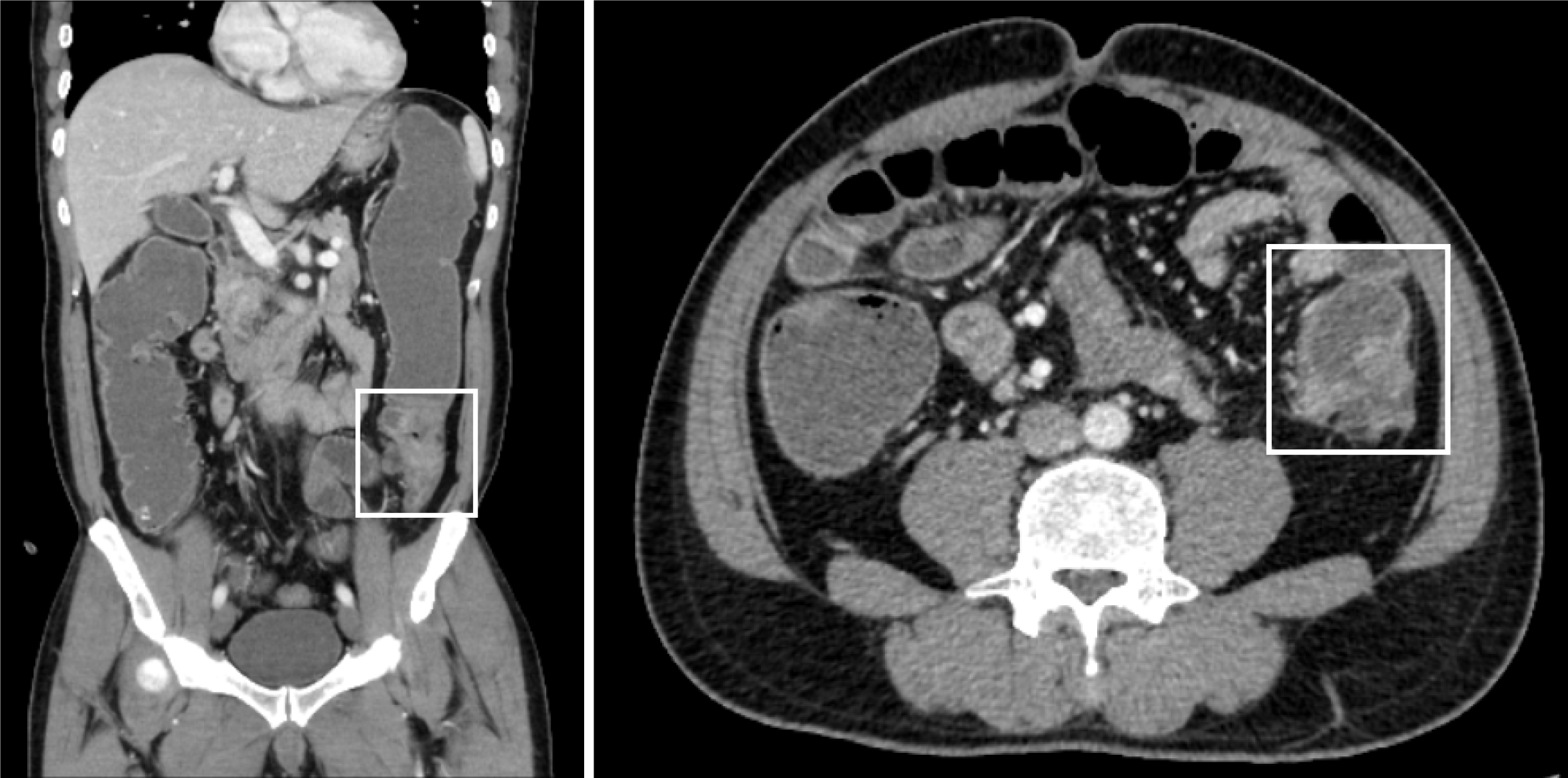
Figure 1 Computed tomography scans of the patient upon admission: The diseased segment exhibited thickening of the intestinal wall, rough surface texture, and significantly increased enhancement on the enhanced scan.
The proximal colon was obstructive dilatation.
- Citation: Wu JC, Cheng HX, Lan QS, Xu HY, Zeng YJ, Lai W, Chu ZH. Penile metastasis from colon cancer with BRAFV600E mutation treated with BRAF/MEK-targeted therapy plus cetuximab: A case report. World J Gastrointest Oncol 2025; 17(3): 100152
- URL: https://www.wjgnet.com/1948-5204/full/v17/i3/100152.htm
- DOI: https://dx.doi.org/10.4251/wjgo.v17.i3.100152