
















©The Author(s) 2025.
World J Gastrointest Oncol. Dec 15, 2025; 17(12): 114390
Published online Dec 15, 2025. doi: 10.4251/wjgo.v17.i12.114390
Published online Dec 15, 2025. doi: 10.4251/wjgo.v17.i12.114390
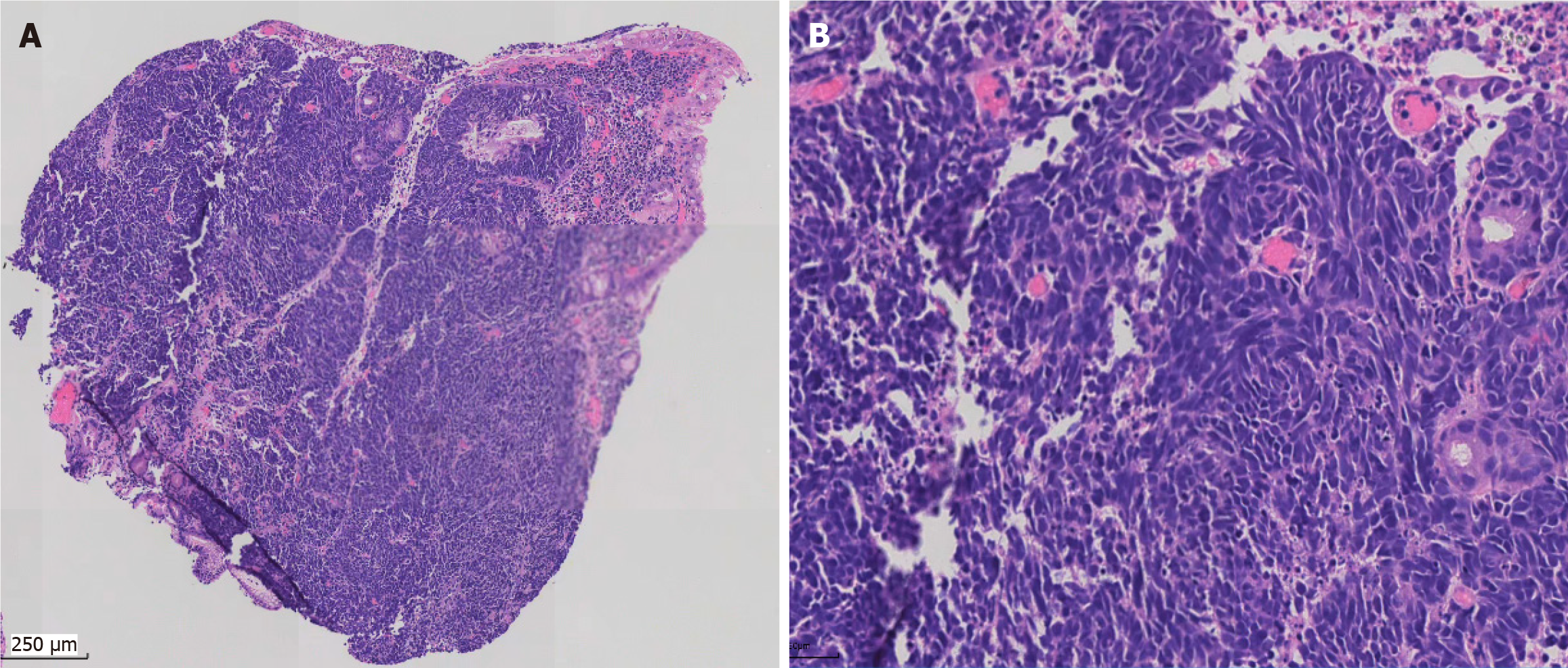
Figure 3 Pathological morphology of gastric small cell carcinoma.
A: Under low-power magnification, sheets of small blue cells infiltrated into the mucosa; B: Under high-power magnification, tumor cells were poorly differentiated, with scant cytoplasm and high mitosis.
- Citation: Zhang XL, Zhang JY, Xie L, Li H, Wang L. Advanced gastric small cell carcinoma with immunotherapy-based treatment: A case report. World J Gastrointest Oncol 2025; 17(12): 114390
- URL: https://www.wjgnet.com/1948-5204/full/v17/i12/114390.htm
- DOI: https://dx.doi.org/10.4251/wjgo.v17.i12.114390