
















©The Author(s) 2025.
World J Gastrointest Oncol. Dec 15, 2025; 17(12): 113524
Published online Dec 15, 2025. doi: 10.4251/wjgo.v17.i12.113524
Published online Dec 15, 2025. doi: 10.4251/wjgo.v17.i12.113524
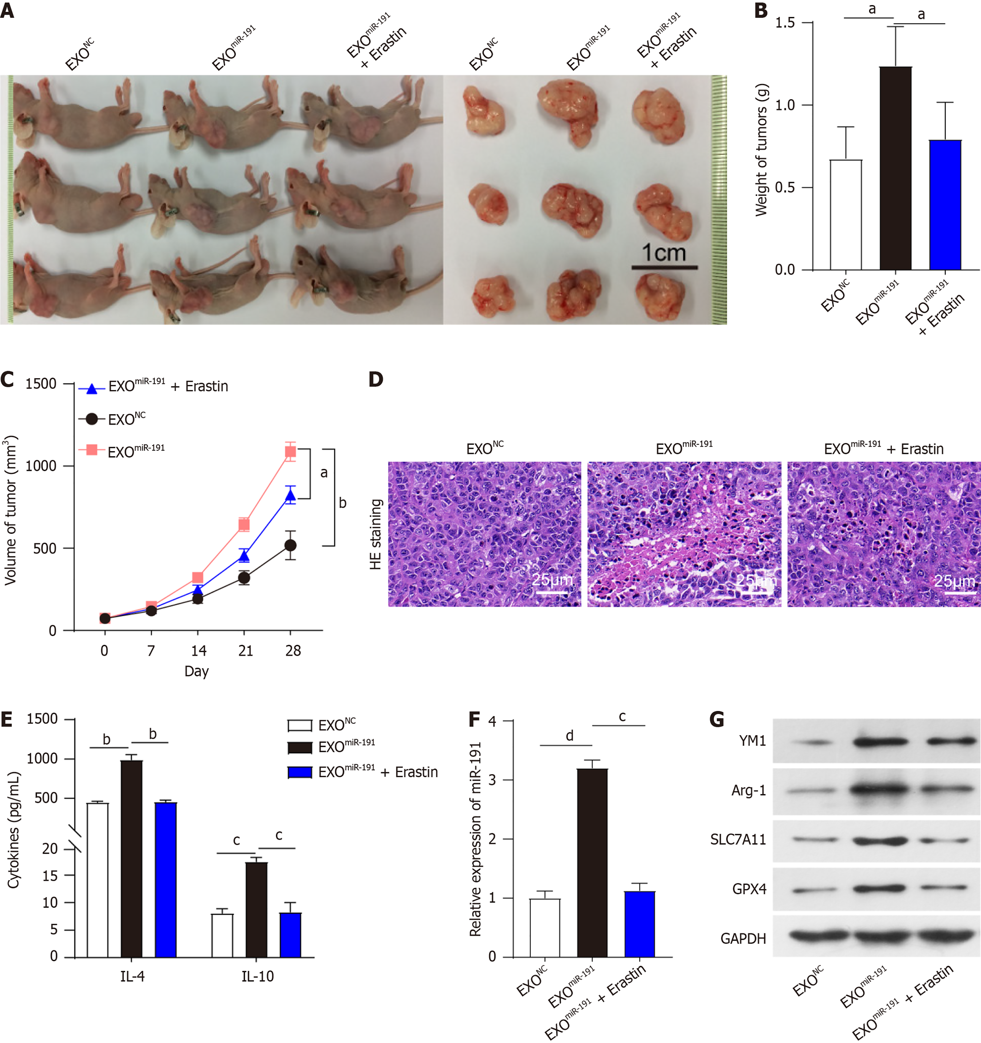
Figure 6 Exosomal miR-191 from colorectal cancer cells promotes growth, and alters pathological structure of subcutaneous tumors in colorectal cancer nude mice by inducing M2 polarization of macrophages.
A: Nude mice were injected subcutaneously with colorectal cancer cells and exosomal miR-191, which were then processed with M2 type macrophage inhibitor erastin; Mice were killed after 28 days, and the tumors were removed; B: Tumor weight was measured; C: Tumor volume was measured; D: Hematoxylin and eosin staining monitored the histopathological changes of the tumors; E: Levels of interleukin (IL)-4 and IL-10 were measured by enzyme-linked immunosorbent assay; F: Expression of miR-191 in tumor tissues was analyzed by quantitative reverse transcription polymerase chain reaction; G: Changes in SLC7A11, GPX4, chitinase-like protein 3 and arginase-1 expression were assessed using western blotting in tumor tissues. aP < 0.05. bP < 0.01. cP < 0.001. dP < 0.0001. Exo: Exosome; NC: Negative control; IL: Interleukin; HE: Hematoxylin and eosin; YM1: Chitinase-like protein 3; Arg-1: Arginase-1; GAPDH: Glyceraldehyde-3-phosphate dehydrogenase.
- Citation: Zhao QY, Wei SJ. Exosomal miR-191 promotes colorectal cancer progression by inducing M2 macrophage polarization and inhibiting ferroptosis. World J Gastrointest Oncol 2025; 17(12): 113524
- URL: https://www.wjgnet.com/1948-5204/full/v17/i12/113524.htm
- DOI: https://dx.doi.org/10.4251/wjgo.v17.i12.113524