
















©The Author(s) 2025.
World J Gastrointest Oncol. Dec 15, 2025; 17(12): 113524
Published online Dec 15, 2025. doi: 10.4251/wjgo.v17.i12.113524
Published online Dec 15, 2025. doi: 10.4251/wjgo.v17.i12.113524
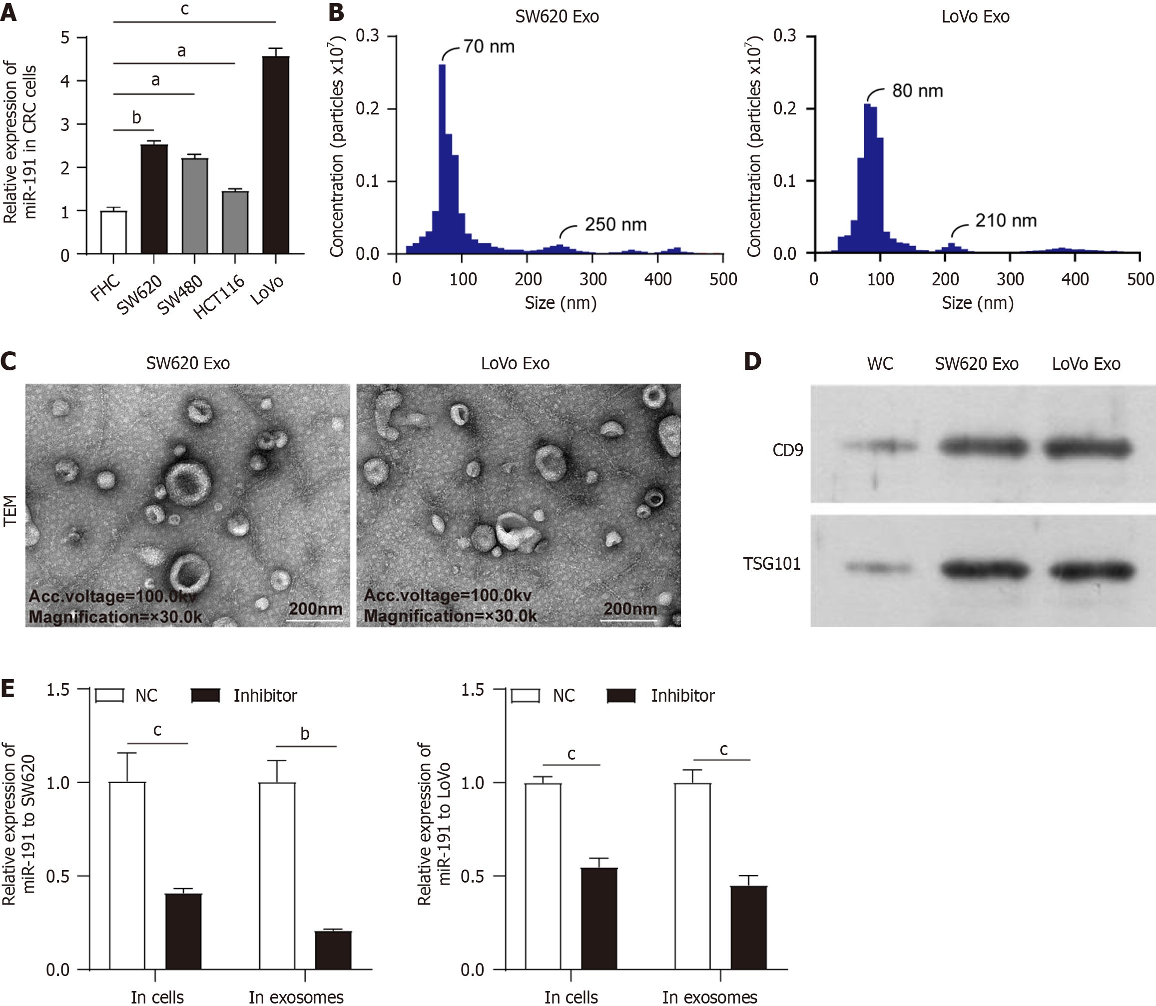
Figure 1 Extraction and identification of supernatant exosomes from colorectal cancer cells after miR-191 inhibition.
A: Quantitative reverse transcription polymerase chain reaction (qRT-PCR) was performed to analyze the expression change of miR-191 in fetal human colon and colorectal cancer cells (SW620, SW480, HCT116 and LoVo); B: Size distribution and number of the isolated exosomes were displayed by nano sight particle-tracking analysis; C: Transmission electron microscopy showed the size and shape of the exosomes derived from SW620 and LoVo cells. Scale bar = 200 nm; D: Expression of cluster of differentiation 9 and TSG101 was confirmed by western blotting in exosomes secreted by SW620 and LoVo cells; E: QRT-PCR analysis of miR-191 expression in SW620- and LoVo-derived exosomes after transfection with miR-191 inhibitor. aP < 0.05. bP < 0.01. cP < 0.001. CRC: Colorectal cancer; FHC: Fetal human colon; Exo: Exosome; TEM: Transmission electron microscopy; CD: Cluster of differentiation; WC: Whole cell lysate; NC: Negative control.
- Citation: Zhao QY, Wei SJ. Exosomal miR-191 promotes colorectal cancer progression by inducing M2 macrophage polarization and inhibiting ferroptosis. World J Gastrointest Oncol 2025; 17(12): 113524
- URL: https://www.wjgnet.com/1948-5204/full/v17/i12/113524.htm
- DOI: https://dx.doi.org/10.4251/wjgo.v17.i12.113524