
















©The Author(s) 2025.
World J Gastrointest Oncol. Dec 15, 2025; 17(12): 113341
Published online Dec 15, 2025. doi: 10.4251/wjgo.v17.i12.113341
Published online Dec 15, 2025. doi: 10.4251/wjgo.v17.i12.113341
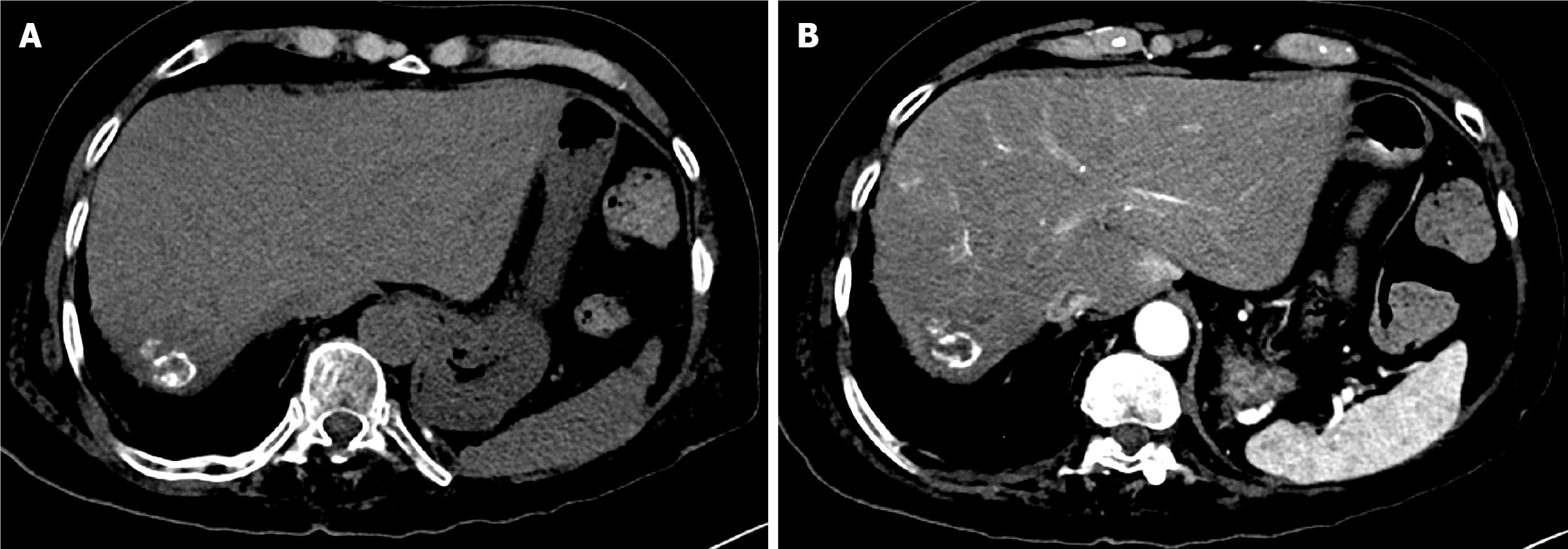
Figure 12 Six-year follow-up computed tomography of the abdomen after the second drug-eluting bead transarterial chemoembolization in Case 2.
A: Pre-contrast computed tomography (CT) demonstrated dense, progressive peripheral concentric calcification surrounding the treated hepatic mass; B: Arterial phase CT showed a non-enhancing mass, consistent with sustained complete response.
- Citation: Alharbi SR. Tumor calcification and sustained complete response after chemoembolization in hepatocellular carcinoma: Two case reports and review of literature. World J Gastrointest Oncol 2025; 17(12): 113341
- URL: https://www.wjgnet.com/1948-5204/full/v17/i12/113341.htm
- DOI: https://dx.doi.org/10.4251/wjgo.v17.i12.113341