
















©The Author(s) 2025.
World J Gastrointest Oncol. Dec 15, 2025; 17(12): 113341
Published online Dec 15, 2025. doi: 10.4251/wjgo.v17.i12.113341
Published online Dec 15, 2025. doi: 10.4251/wjgo.v17.i12.113341
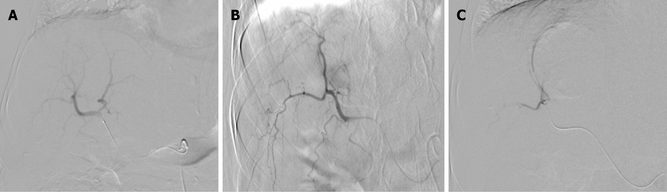
Figure 3 Digital subtraction angiography images during the first drug-eluting bead transarterial chemoembolization session in Case 1.
A: Right hepatic artery non-selective angiogram; B: Selective hepatic angiogram showed a hypervascular mass; C: Superselective angiogram demonstrated complete stasis of the vascular supply of the tumor after embolization.
- Citation: Alharbi SR. Tumor calcification and sustained complete response after chemoembolization in hepatocellular carcinoma: Two case reports and review of literature. World J Gastrointest Oncol 2025; 17(12): 113341
- URL: https://www.wjgnet.com/1948-5204/full/v17/i12/113341.htm
- DOI: https://dx.doi.org/10.4251/wjgo.v17.i12.113341