
















©The Author(s) 2025.
World J Gastrointest Oncol. Dec 15, 2025; 17(12): 113289
Published online Dec 15, 2025. doi: 10.4251/wjgo.v17.i12.113289
Published online Dec 15, 2025. doi: 10.4251/wjgo.v17.i12.113289
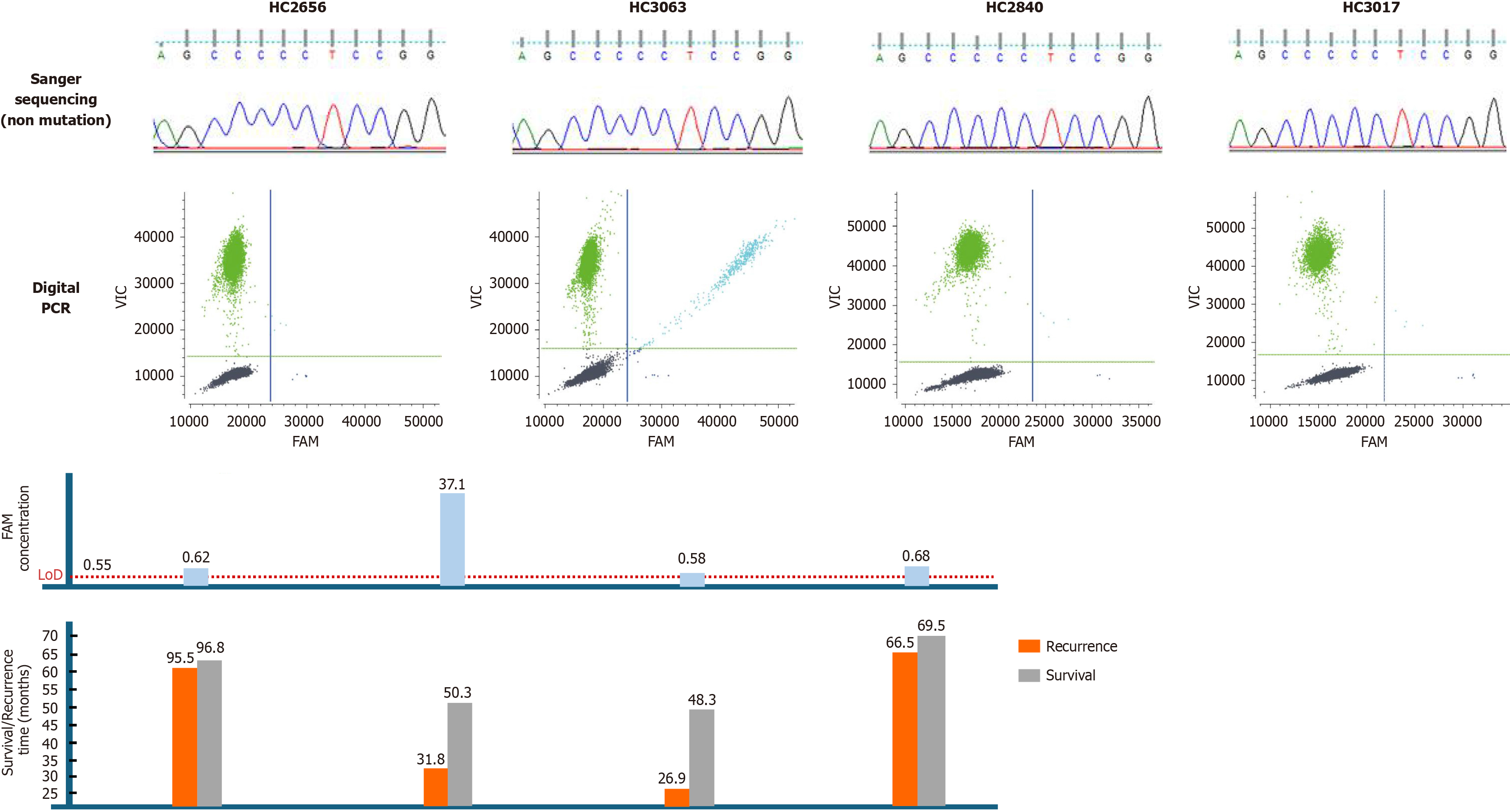
Figure 4 Illustration of sequencing results and follow-up data in four samples, which were positive for telomerase reverse transcriptase C228T by digital polymerase chain reaction assay, but were all negative by Sanger sequencing.
Each column represents a sample of one patient with hepatocellular carcinoma after surgical resection. At the top of the figure, Sanger sequencing results (all were non mutated); In the middle of the figure, the two-dimensional digital polymerase chain reaction plot (the color of line: Green, wild-type; dark blue, mutant type; light blue, droplets with mutant and wild-type allele; dark grey, negative droplets) and the concentration (cp/μL) of the telomerase reverse transcriptase C228T mutant allele targeted by the 6-carboxyfluorescein dye labeled probes. 6-carboxyfluorescein concentration higher than LoD was positive. Clinical follow-up data collected from each patient are shown at the bottom of the figure. The orange and grey bars represent the time from surgery to tumor recurrence and death, respectively. PCR: Polymerase chain reaction.
- Citation: Aizimuaji Z, Hu N, Li HY, Wang XJ, Ma S, Wang YR, Zheng RQ, Li Z, Zhao H, Rong WQ, Xiao T. Optimized digital polymerase chain reaction enables detection of telomerase reverse transcriptase C228T mutation for prognostic assessment in hepatocellular carcinoma. World J Gastrointest Oncol 2025; 17(12): 113289
- URL: https://www.wjgnet.com/1948-5204/full/v17/i12/113289.htm
- DOI: https://dx.doi.org/10.4251/wjgo.v17.i12.113289