
















©The Author(s) 2025.
World J Gastrointest Oncol. Dec 15, 2025; 17(12): 113289
Published online Dec 15, 2025. doi: 10.4251/wjgo.v17.i12.113289
Published online Dec 15, 2025. doi: 10.4251/wjgo.v17.i12.113289
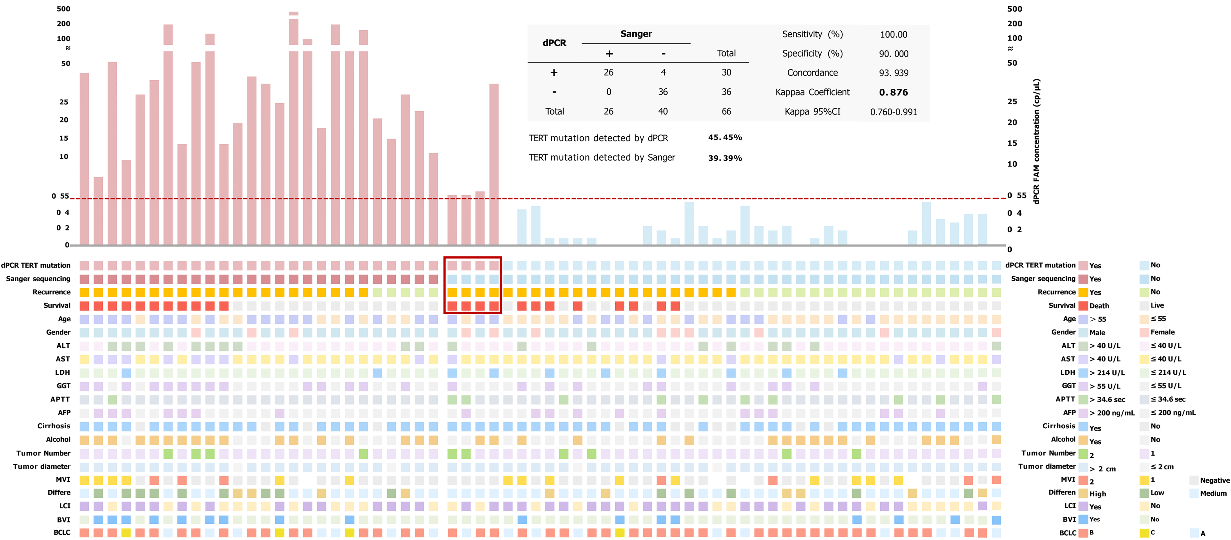
Figure 3 Altered landscape of hepatocellular carcinoma cohort samples, combined with clinical variables (the lower half) and a bar graph of 6-carboxyfluorescein concentration (the upper half) of telomerase reverse transcriptase C228T mutation.
A minimum concentration of 0.55 cp/μL (limit of detection) was evaluated to be positive in digital polymerase chain reaction. Comparison of telomerase reverse transcriptase status detected by Sanger sequencing and digital polymerase chain reaction in our hepatocellular carcinoma cohort (the table at the top of this picture). +: Telomerase reverse transcriptase C228T mutated; -: Telomerase reverse transcriptase C228T non-mutated; dPCR: Digital polymerase chain reaction; TERT: Telomerase reverse transcriptase; ALT: Alanine aminotransferase; AST: Aspartate aminotransferase; LDH: Lactate dehydrogenase; GGT: Gamma-glutamyl transferase; APTT: Activated partial thromboplastin time; AFP: Alpha fetoprotein; MVI: Microvascular invasion; LCI: Liver capsular invasion; BVI: Blood vessel invasion; BCLC: Barcelona Clinic Liver Cancer; CI: Confidence interval.
- Citation: Aizimuaji Z, Hu N, Li HY, Wang XJ, Ma S, Wang YR, Zheng RQ, Li Z, Zhao H, Rong WQ, Xiao T. Optimized digital polymerase chain reaction enables detection of telomerase reverse transcriptase C228T mutation for prognostic assessment in hepatocellular carcinoma. World J Gastrointest Oncol 2025; 17(12): 113289
- URL: https://www.wjgnet.com/1948-5204/full/v17/i12/113289.htm
- DOI: https://dx.doi.org/10.4251/wjgo.v17.i12.113289