
















©The Author(s) 2025.
World J Gastrointest Oncol. Dec 15, 2025; 17(12): 113198
Published online Dec 15, 2025. doi: 10.4251/wjgo.v17.i12.113198
Published online Dec 15, 2025. doi: 10.4251/wjgo.v17.i12.113198
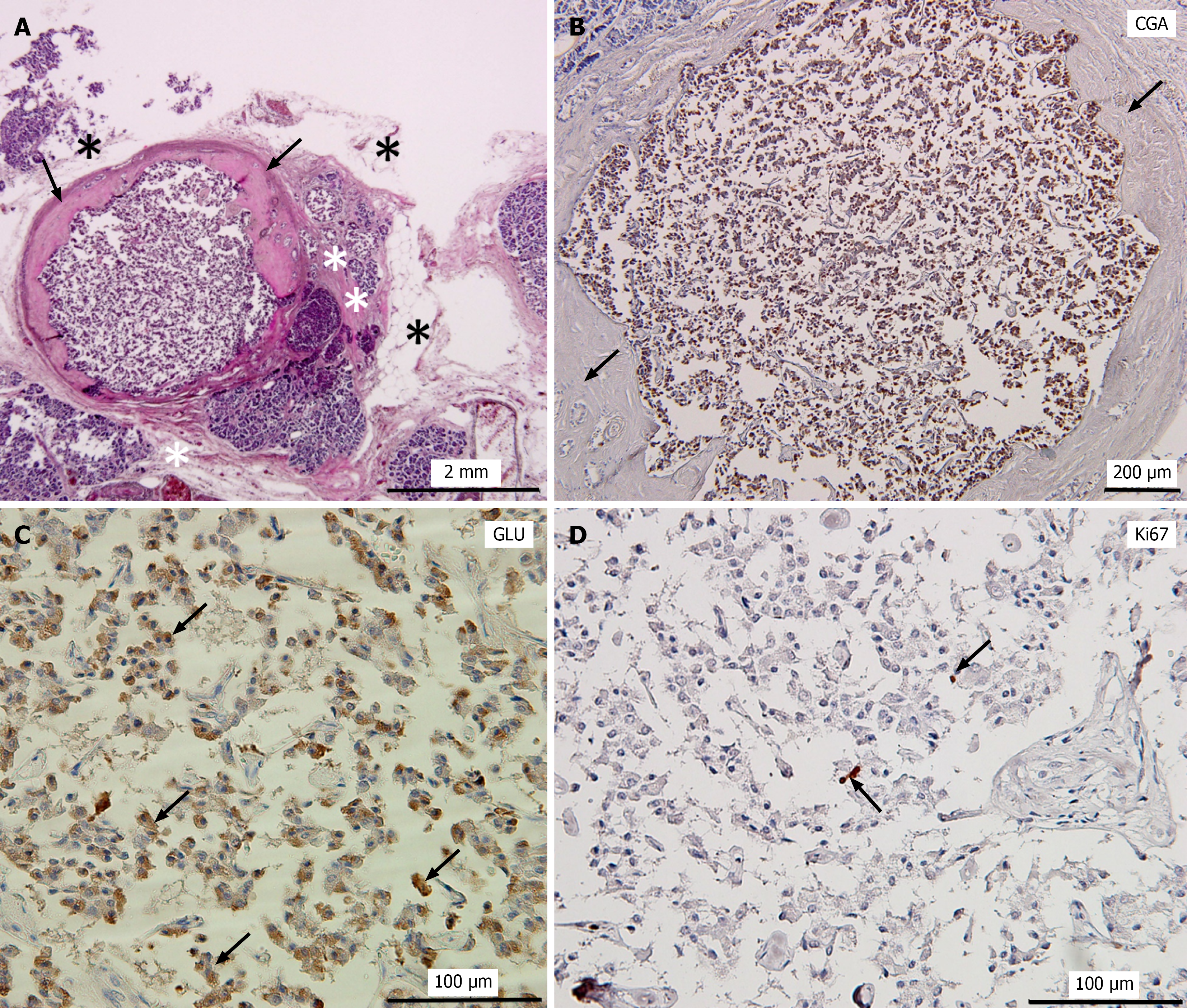
Figure 3 Histopathological changes of pancreatic neuroendocrine microtumors in Case 5.
A: Hematoxylin and eosin staining of pancreatic neuroendocrine microtumors (PNEMTs) of Case 5; B: Immunostaining of PNEMTs in A with chromogranin A antibody; C: Immunostaining of PNEMTs with glucagon (GLU) antibody; D: Immunostaining of PNEMTs with Ki-67 antibody. Black arrows in A and B: Indicate that the outer layer of the tumor was surrounded by a thick fibrotic wall; Black asterisk in A: Fatty degeneration; white two asterisks in A: Fibrosis; Black arrows in C: GLU-positive PNEMTs cells; Black arrows in D: A small number of Ki-67-positive PNEMTs cells. CGA: Chromogranin A; GLU: Glucagon.
- Citation: Yang T, Ren K, Chen XQ, Toriumi T, Natsuyama Y, Li J, Sukeda A, Nagao T, Yi SQ. Pancreatic neuroendocrine microtumors in the elderly: A retrospective study using cadaveric pancreatic tissue. World J Gastrointest Oncol 2025; 17(12): 113198
- URL: https://www.wjgnet.com/1948-5204/full/v17/i12/113198.htm
- DOI: https://dx.doi.org/10.4251/wjgo.v17.i12.113198