
















©The Author(s) 2025.
World J Gastrointest Oncol. Dec 15, 2025; 17(12): 112853
Published online Dec 15, 2025. doi: 10.4251/wjgo.v17.i12.112853
Published online Dec 15, 2025. doi: 10.4251/wjgo.v17.i12.112853
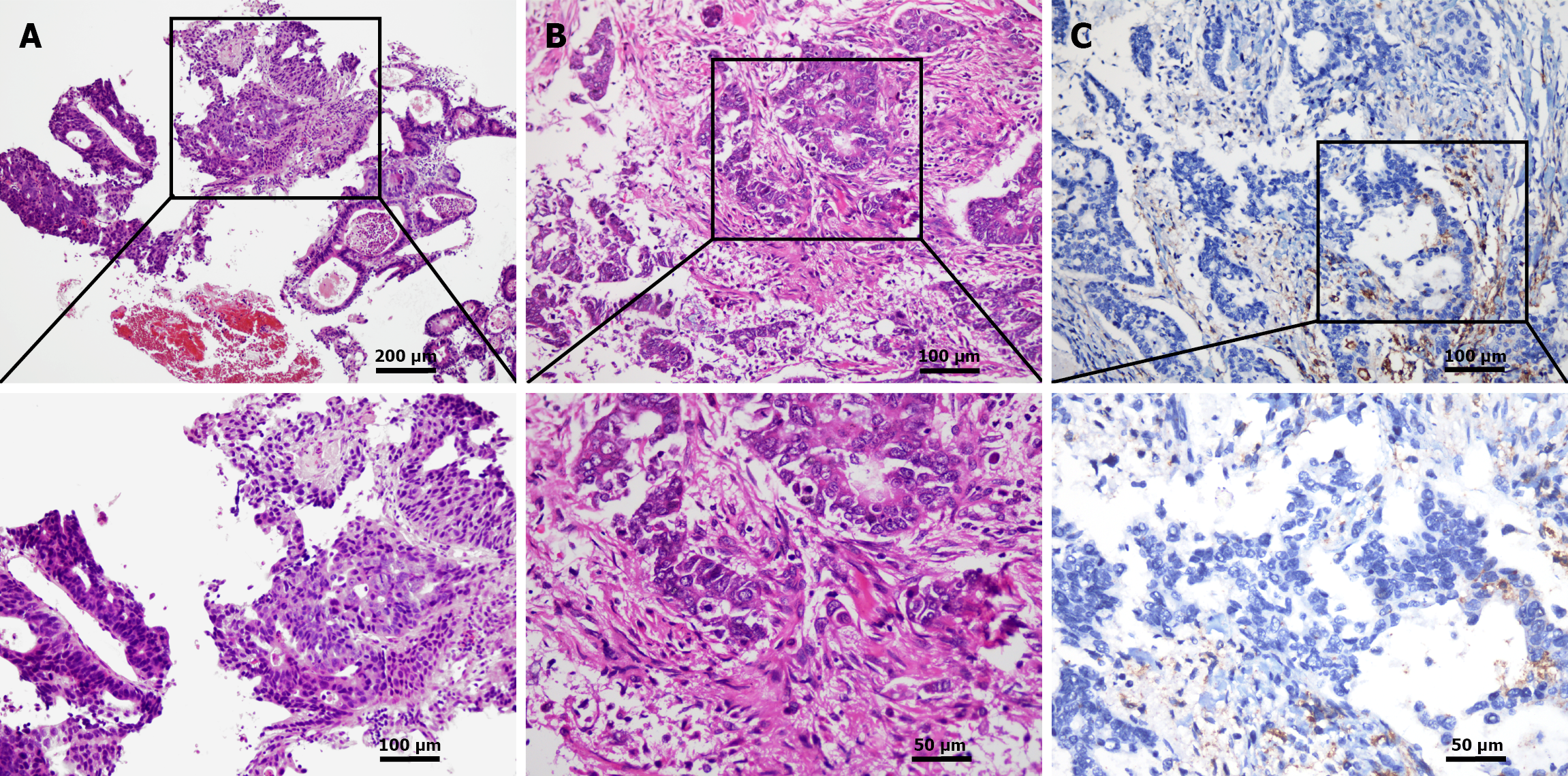
Figure 1 Pathology and immunohistochemistry of the endoscopic biopsy and resected specimen.
A: Hematoxylin and eosin (HE) staining of the endoscopic biopsy showed suggestive of gastric cancer: Moderately differentiated adenocarcinoma, HE × 100 and HE × 200; B: HE staining of the resected specimen confirms the diagnosis of gastric cancer: Moderately to poorly differentiated adenocarcinoma, with infiltration through the entire thickness of the gastric wall into the serosal fibrous adipose tissue, HE × 200 and HE × 400; C: Immunohistochemistry (IHC) of the resected specimen showed programmed death ligand 1 positivity, IIHC × 200 and IHC × 400.
- Citation: Pan J, Li P, Zhou YH, Pan TT, Chen YT, Chu XY. Long-term survival after treatment of gastric cancer with S-1 plus oxaliplatin regimen and sintilimab: A case report. World J Gastrointest Oncol 2025; 17(12): 112853
- URL: https://www.wjgnet.com/1948-5204/full/v17/i12/112853.htm
- DOI: https://dx.doi.org/10.4251/wjgo.v17.i12.112853