
















©The Author(s) 2025.
World J Gastrointest Oncol. Dec 15, 2025; 17(12): 112366
Published online Dec 15, 2025. doi: 10.4251/wjgo.v17.i12.112366
Published online Dec 15, 2025. doi: 10.4251/wjgo.v17.i12.112366
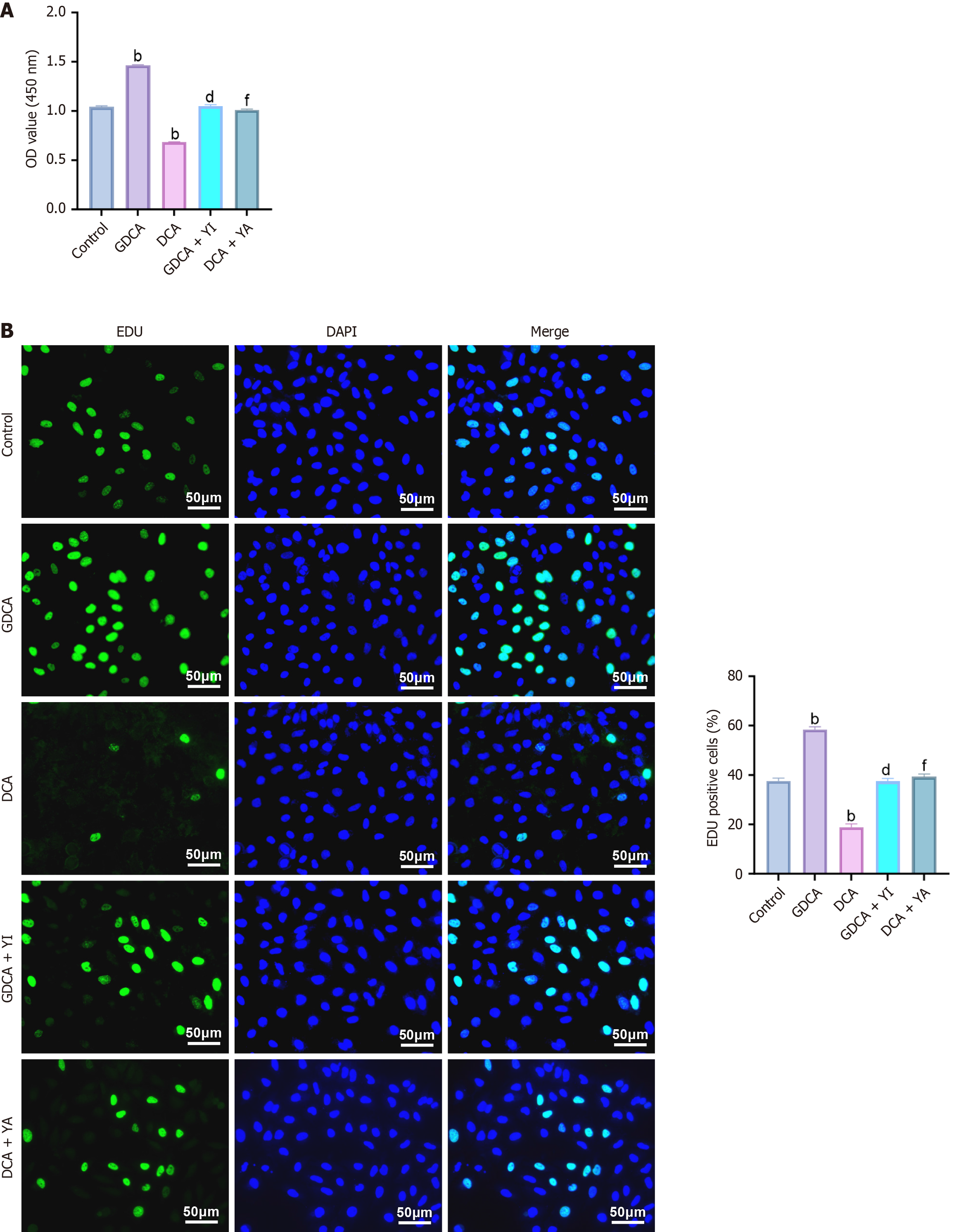
Figure 2 Effects of glycochenodeoxycholic acid and deoxycholic acid on cholangiocarcinoma cell proliferation.
A: Cell Counting Kit-8 was used to detect the viability of HuCCT1 cells in the control, glycochenodeoxycholic acid (GDCA), deoxycholic acid (DCA), GDCA + YI, and DCA + YI groups; B: 5-ethynyl-2’-deoxyuridine staining was used to detect the DNA replication ability of HuCCT1 cells in the control, GDCA, DCA, GDCA + YI, and DCA + YA groups. Statistical analysis was conducted using one-way ANOVA with Tukey’s post hoc test. Significance is indicated as: bP < 0.01 vs control; dP < 0.01 vs GDCA; fP < 0.01 vs DCA. Data are presented as mean ± SD (n = 3 independent experiments). OD: Optical density; DAPI: 4’,6-diamidino-2-phenylindole; EDU: 5-ethynyl-2’-deoxyuridine; GDCA: Glycochenodeoxycholic acid; DCA: Deoxycholic acid.
- Citation: Hu J, Zhang G, Yang S, Shen XF, Zhou M, Huang LS, Lan HM. Involvement of bile acids in cholangiocarcinoma progression via the Hippo-yes-associated protein signaling pathway. World J Gastrointest Oncol 2025; 17(12): 112366
- URL: https://www.wjgnet.com/1948-5204/full/v17/i12/112366.htm
- DOI: https://dx.doi.org/10.4251/wjgo.v17.i12.112366