
















©The Author(s) 2025.
World J Gastrointest Oncol. Nov 15, 2025; 17(11): 112203
Published online Nov 15, 2025. doi: 10.4251/wjgo.v17.i11.112203
Published online Nov 15, 2025. doi: 10.4251/wjgo.v17.i11.112203
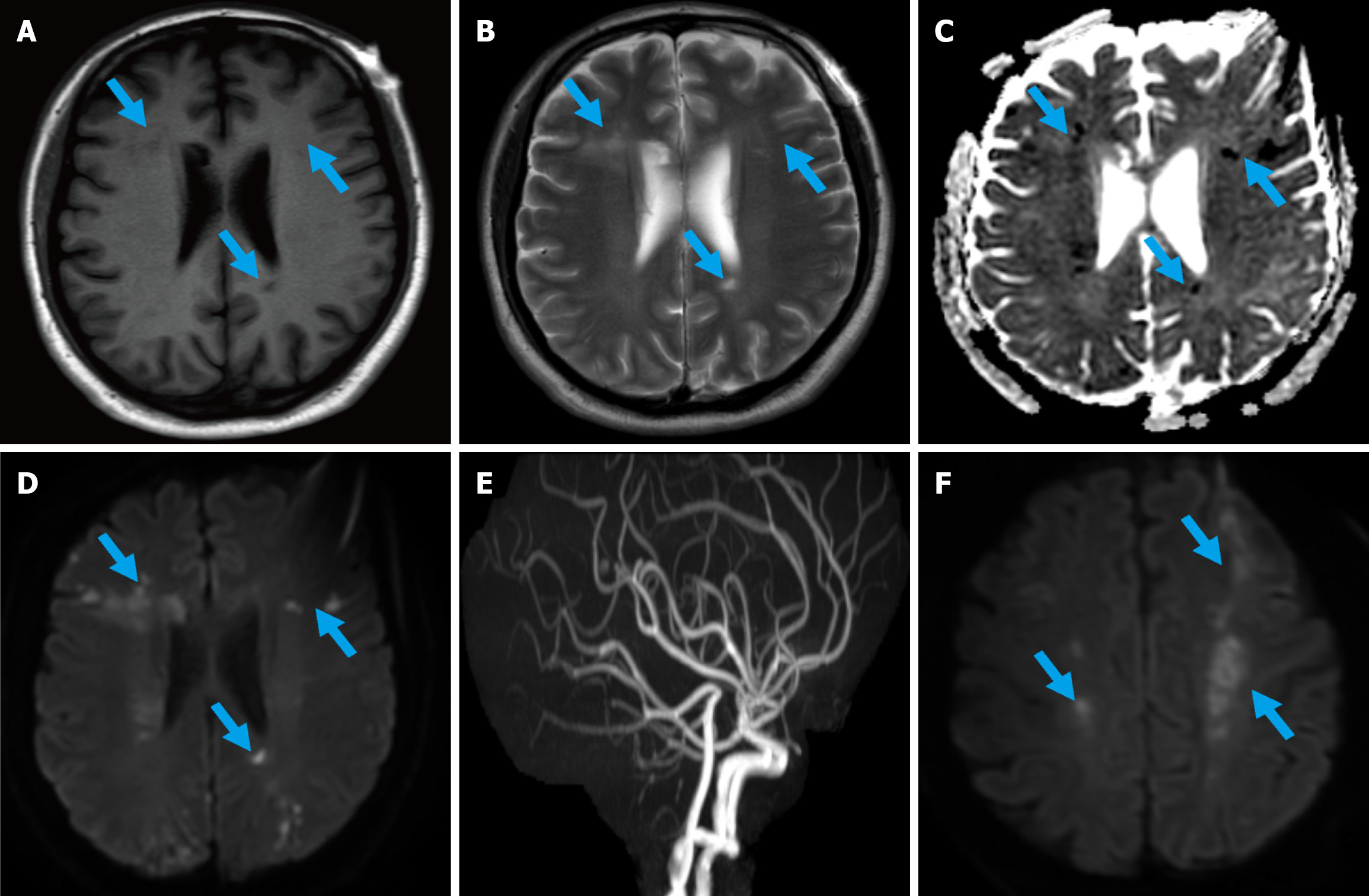
Figure 3 Brain magnetic resonance imaging and magnetic resonance angiography images are presented.
A-D: Bilateral multiple paraventricular infarctions are demonstrated on T1-weighted images, T2-weighted images, apparent diffusion coefficient mapping, and diffusion-weighted imaging (DWI) sequences; E: Magnetic resonance angiography showing no significant abnormalities; F: Follow-up DWI revealing newly developed infarct lesions in the bilateral parietal and frontal lobes.
- Citation: Wang QY, Xia WH, Wan W, Liu JP. Pancreatic cancer initially presenting with acute renal infarction: A case report. World J Gastrointest Oncol 2025; 17(11): 112203
- URL: https://www.wjgnet.com/1948-5204/full/v17/i11/112203.htm
- DOI: https://dx.doi.org/10.4251/wjgo.v17.i11.112203