
















©The Author(s) 2025.
World J Gastrointest Oncol. Nov 15, 2025; 17(11): 112089
Published online Nov 15, 2025. doi: 10.4251/wjgo.v17.i11.112089
Published online Nov 15, 2025. doi: 10.4251/wjgo.v17.i11.112089
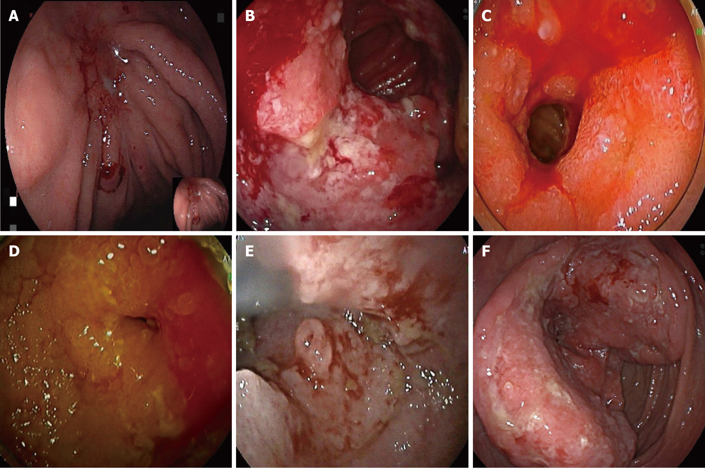
Figure 3 Endoscopic features of lymphoma.
A: An ulcer was observed at the junction of the gastric fundus and body, with a white coating in the center. The surrounding mucosa exhibited congestion, edema, and convergence, with a rigid sensation upon insufflation, poor peristalsis, and slight oozing; B: A circumferential ulcerative and proliferative lesion was found at approximately 300 cm of the small intestine, with an uneven surface covered by dirty coating, leading to intestinal stenosis; C: A circumferential ulcerative mass causing luminal stenosis was detected 160 cm from the pylorus in the small intestine. The surrounding mucosa was congested and reddened, with local mucosal rigidity and friability, making it prone to bleeding; D: A circumferential ulcerative and proliferative lesion was observed at approximately 300 cm of the small intestine via anal approach, with an uneven surface covered by dirty coating, leading to intestinal stenosis; E: A mucosal mass was found 50 cm from the ileocecal junction in the small intestine, with surface erosion and fresh blood stains. Mild luminal stenosis was noted at the lesion site; F: A circumferential space-occupying lesion was detected in the ascending colon during endoscopy, showing surface ulceration and a hard texture.
- Citation: Yang CX, Xu LX, Liu J, Qiao HL, Dong ZW, Jiang D, Gu GL. Clinicopathological characteristics and surgical value of primary gastrointestinal lymphoma. World J Gastrointest Oncol 2025; 17(11): 112089
- URL: https://www.wjgnet.com/1948-5204/full/v17/i11/112089.htm
- DOI: https://dx.doi.org/10.4251/wjgo.v17.i11.112089