
















©The Author(s) 2025.
World J Gastrointest Oncol. Nov 15, 2025; 17(11): 112089
Published online Nov 15, 2025. doi: 10.4251/wjgo.v17.i11.112089
Published online Nov 15, 2025. doi: 10.4251/wjgo.v17.i11.112089
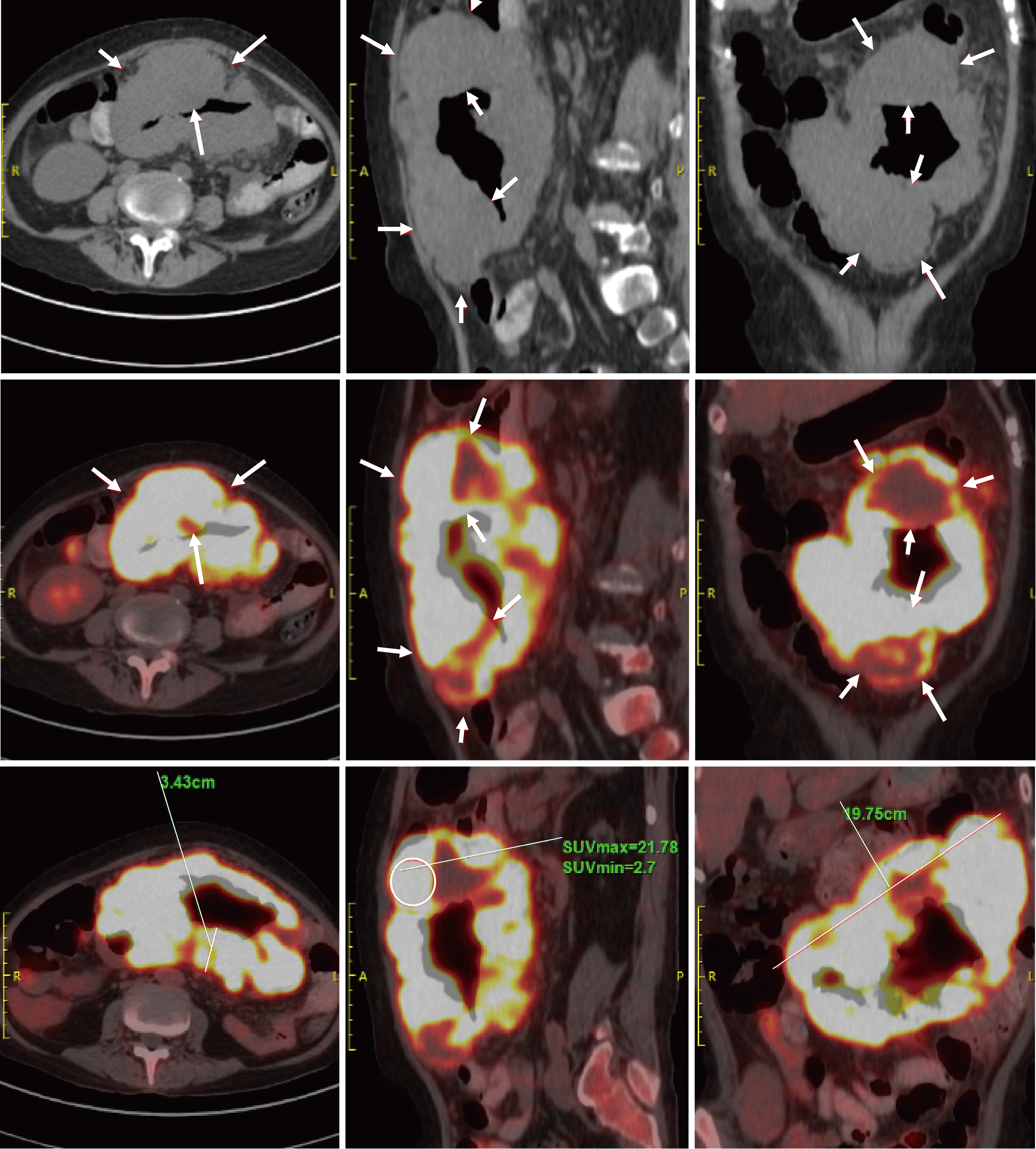
Figure 2 Positron emission tomography/computed tomography findings.
Segmental circumferential thickening and mass formation were noted in the second segment of the small intestine, extending from the left upper to mid-abdomen, with significantly increased fluorodeoxyglucose uptake. Multiple enlarged lymph nodes were observed in the abdominal cavity surrounding the small intestinal lesion, also demonstrating markedly elevated fluorodeoxyglucose uptake. Arrow: Segmental small bowel wall thickening and a mass, respectively. The circled areas highlight regions with increased fluorodeoxyglucose uptake. The solid lines mark the measured lengths.
- Citation: Yang CX, Xu LX, Liu J, Qiao HL, Dong ZW, Jiang D, Gu GL. Clinicopathological characteristics and surgical value of primary gastrointestinal lymphoma. World J Gastrointest Oncol 2025; 17(11): 112089
- URL: https://www.wjgnet.com/1948-5204/full/v17/i11/112089.htm
- DOI: https://dx.doi.org/10.4251/wjgo.v17.i11.112089