
















©The Author(s) 2025.
World J Gastrointest Oncol. Nov 15, 2025; 17(11): 112089
Published online Nov 15, 2025. doi: 10.4251/wjgo.v17.i11.112089
Published online Nov 15, 2025. doi: 10.4251/wjgo.v17.i11.112089
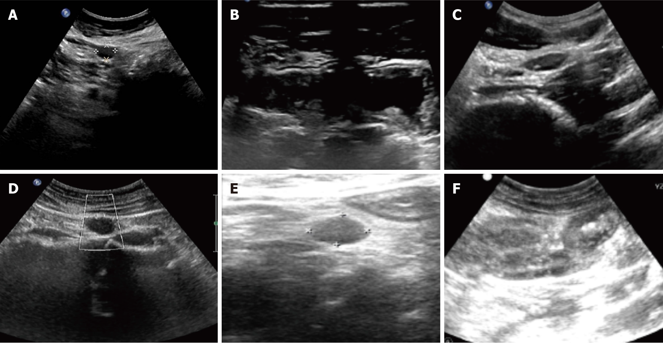
Figure 1 Ultrasonographic findings.
A: Multiple hypoechoic lesions are visualized in the peritoneal cavity. The largest, located in the perigastric region, measures 1.5 cm × 1.1 cm, demonstrates well-defined borders and a regular morphology, and exhibits homogeneous, markedly hypoechoic internal echogenicity; B: Localized thickening of the intestinal wall was observed in the terminal ileum, with a maximum thickness of approximately 0.5 cm; the layered structure of the wall remained relatively clear; C: Multiple lymph node echoes were detected in the retroperitoneum, with the largest measuring approximately 2.3 cm × 1.0 cm. These lymph nodes had clear boundaries and a regular shape; D: Lymph node echo was observed in the retroperitoneum, with the larger one located at the umbilical level in the right mid-abdomen, measuring 15 cm × 2.2 cm. The lymph node had clear boundaries and a regular shape, but the hilum structure was unclear; E: Multiple hypoechoic areas were found in the intestinal interspace, with the largest measuring 0.7 cm × 1.0 cm; F: Multiple enlarged lymph node echoes were detected in the mesentery, with the largest measuring approximately 0.8 cm × 1.8 cm. These lymph nodes exhibited relatively clear boundaries, an irregular shape, and an unclear hilum structure.
- Citation: Yang CX, Xu LX, Liu J, Qiao HL, Dong ZW, Jiang D, Gu GL. Clinicopathological characteristics and surgical value of primary gastrointestinal lymphoma. World J Gastrointest Oncol 2025; 17(11): 112089
- URL: https://www.wjgnet.com/1948-5204/full/v17/i11/112089.htm
- DOI: https://dx.doi.org/10.4251/wjgo.v17.i11.112089