
















©The Author(s) 2025.
World J Gastrointest Oncol. Nov 15, 2025; 17(11): 110715
Published online Nov 15, 2025. doi: 10.4251/wjgo.v17.i11.110715
Published online Nov 15, 2025. doi: 10.4251/wjgo.v17.i11.110715
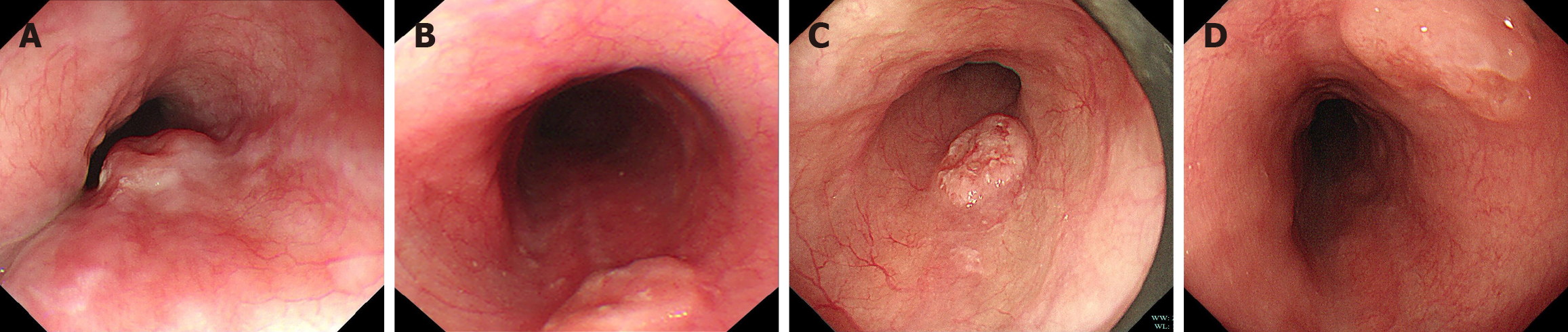
Figure 1 White light endoscopic images of cases 1 to 4.
A: Case 1, a 1.0 cm mucosal elevation with central depression was observed 30 cm from the incisors. The lesion was mobile on palpation; B: Case 2, a 1.2 cm lesion was noted 27 cm from the incisors; C: Case 3, a type 2A mucosal elevation, measuring approximately 1.2 cm × 0.7 cm, was observed 28 cm from the incisors. The lesion exhibited surface erythema, pallor, irregular granularity, erosion, spontaneous bleeding, and well-defined margins; D: Case 4, a mucosal elevation measuring approximately 1.1 cm with slight central depression was detected 30 cm from the incisors.
- Citation: Jin T, Zhou YW, Sun PS, Huang Y, Gao JG, Jin X. Unraveling the characteristics of early esophageal neuroendocrine carcinoma using multi-model endoscopy: A retrospective study of serial cases. World J Gastrointest Oncol 2025; 17(11): 110715
- URL: https://www.wjgnet.com/1948-5204/full/v17/i11/110715.htm
- DOI: https://dx.doi.org/10.4251/wjgo.v17.i11.110715