
















©The Author(s) 2025.
World J Gastrointest Oncol. Nov 15, 2025; 17(11): 110468
Published online Nov 15, 2025. doi: 10.4251/wjgo.v17.i11.110468
Published online Nov 15, 2025. doi: 10.4251/wjgo.v17.i11.110468
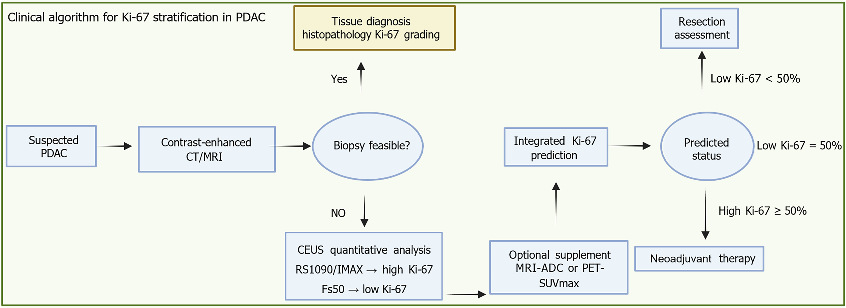
Figure 3 Integrated imaging biomarker pathway for Ki-67 prediction and treatment allocation in biopsy-contraindicated pancreatic ductal adenocarcinoma.
This clinical workflow outlines a decision-making pathway for suspected pancreatic ductal adenocarcinoma, integrating imaging biomarkers to support personalized treatment planning. Following initial contrast-enhanced computed tomography or magnetic resonance imaging for anatomical staging, the feasibility of tissue biopsy is assessed. If biopsy is possible, histopathological Ki-67 grading provides a direct evaluation of tumor proliferative activity. When biopsy is not feasible - such as in patients on anticoagulation therapy or with poor clinical condition - noninvasive contrast-enhanced ultrasound becomes essential. Quan
- Citation: Wu YX, Tian R, Li XW, Guo JY, Tang JF, Zhou CF. Emerging non-invasive imaging biomarkers of Ki-67 in pancreatic cancer: Toward predictive precision oncology. World J Gastrointest Oncol 2025; 17(11): 110468
- URL: https://www.wjgnet.com/1948-5204/full/v17/i11/110468.htm
- DOI: https://dx.doi.org/10.4251/wjgo.v17.i11.110468