















©The Author(s) 2025.
World J Gastrointest Oncol. Nov 15, 2025; 17(11): 110468
Published online Nov 15, 2025. doi: 10.4251/wjgo.v17.i11.110468
Published online Nov 15, 2025. doi: 10.4251/wjgo.v17.i11.110468
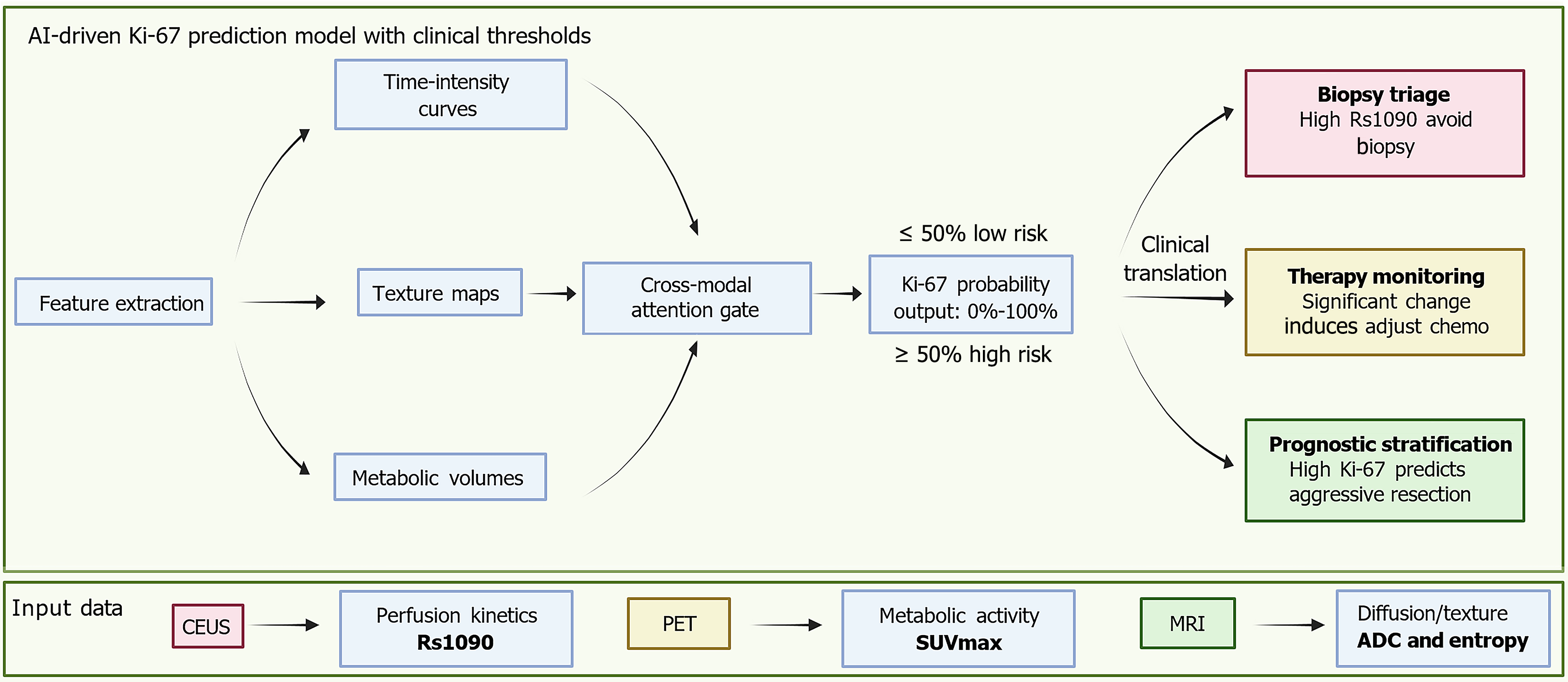
Figure 2 Unified predictive model for Ki-67 using multimodal imaging.
This schematic presents an artificial intelligence fusion model that integrates multimodal imaging data. These include contrast-enhanced ultrasound derived perfusion kinetics, specifically rise slope 10%-90%, positron emission tomography based metabolic activity represented by the maximum standardized uptake value, and magnetic resonance imaging derived diffusion and texture metrics such as apparent diffusion coefficient and entropy. The model extracts key imaging features including time-intensity curves, metabolic volumes, and texture maps, which are then synthesized using a cross-modal attention gate to generate a probabilistic Ki-67 output ranging from 0%-100%. Clinically, this artificial intelligence-derived output supports three major decisions. First, biopsy triage, where a high rise slope 10%-90% value may help avoid invasive procedures in patients at risk for biopsy-related complications. Second, therapy monitoring, where significant changes in perfusion parameters or rising maximum standardized uptake values during neoadjuvant treatment can prompt early adjustments to chemotherapy. Third, prognostic stratification, where high-probability Ki-67 spatial predictions assist in identifying aggressive tumor regions and support surgical margin planning. AI: Artificial intelligence; Rs1090: Rise slope 10%-90%; CEUS: Contrast-enhanced ultrasound; PET: Positron emission tomography; SUVmax: Maximum standardized uptake value; MRI: Magnetic resonance imaging; ADC: Apparent diffusion coefficient.
- Citation: Wu YX, Tian R, Li XW, Guo JY, Tang JF, Zhou CF. Emerging non-invasive imaging biomarkers of Ki-67 in pancreatic cancer: Toward predictive precision oncology. World J Gastrointest Oncol 2025; 17(11): 110468
- URL: https://www.wjgnet.com/1948-5204/full/v17/i11/110468.htm
- DOI: https://dx.doi.org/10.4251/wjgo.v17.i11.110468