
















©The Author(s) 2025.
World J Gastrointest Oncol. Oct 15, 2025; 17(10): 109506
Published online Oct 15, 2025. doi: 10.4251/wjgo.v17.i10.109506
Published online Oct 15, 2025. doi: 10.4251/wjgo.v17.i10.109506
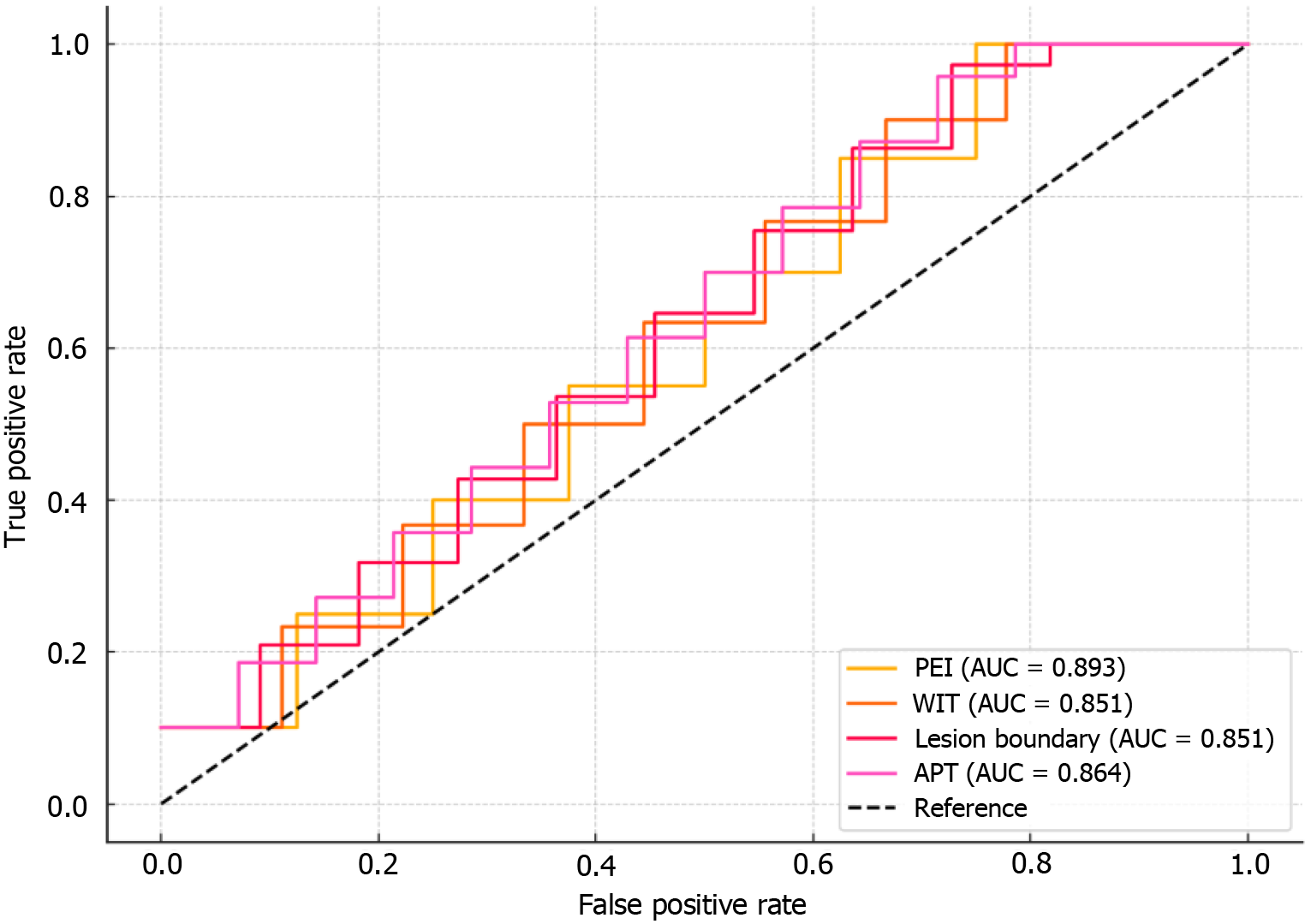
Figure 4 Receiver operating characteristic curve of each indicator.
PEI: Peak enhancement intensity; WIT: Washout initiation time; APT: Arterial phase enhancement time; AUC: Area under the curve.
- Citation: Dong Y, Chen LP, He JG, Yang QQ, Hu ZW. Clinical value of contrast-enhanced ultrasound in early diagnosis of hepatocellular carcinoma. World J Gastrointest Oncol 2025; 17(10): 109506
- URL: https://www.wjgnet.com/1948-5204/full/v17/i10/109506.htm
- DOI: https://dx.doi.org/10.4251/wjgo.v17.i10.109506