
















©The Author(s) 2025.
World J Gastrointest Oncol. Oct 15, 2025; 17(10): 109450
Published online Oct 15, 2025. doi: 10.4251/wjgo.v17.i10.109450
Published online Oct 15, 2025. doi: 10.4251/wjgo.v17.i10.109450
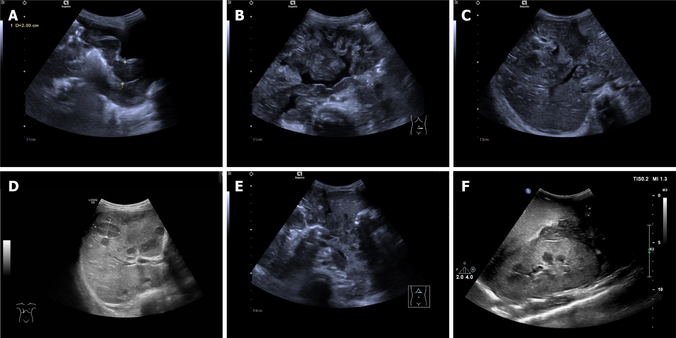
Figure 3 Manifestations of abdominal cavity and organ invasion in intestinal lymphoma.
A: The intestinal wall (sigmoid colon and rectum) of the primary part was circular thickening thickened, with decreased echo and small intestinal lumen; B: In the remaining abdominal cavities, the intestinal wall was widely thickened, and the thickening was not as obvious as that of the primary lesion, with interintestinal effusion; C and D: Extremely hypoechoic distributed along the Greenland sheath in the liver and multiple hypoechoic metastatic foci in the liver; E: A widely distributed low-echo area was observed around the pancreas; F: Scattered low-echo areas in the kidney.
- Citation: Huang SF, Yang F, Chen WJ, Zhang XH. Ultrasound features of primary intestinal lymphoma in children and their correlation with prognosis: A two-center experiment. World J Gastrointest Oncol 2025; 17(10): 109450
- URL: https://www.wjgnet.com/1948-5204/full/v17/i10/109450.htm
- DOI: https://dx.doi.org/10.4251/wjgo.v17.i10.109450