
















©The Author(s) 2024.
World J Gastrointest Oncol. Jul 15, 2024; 16(7): 3341-3349
Published online Jul 15, 2024. doi: 10.4251/wjgo.v16.i7.3341
Published online Jul 15, 2024. doi: 10.4251/wjgo.v16.i7.3341
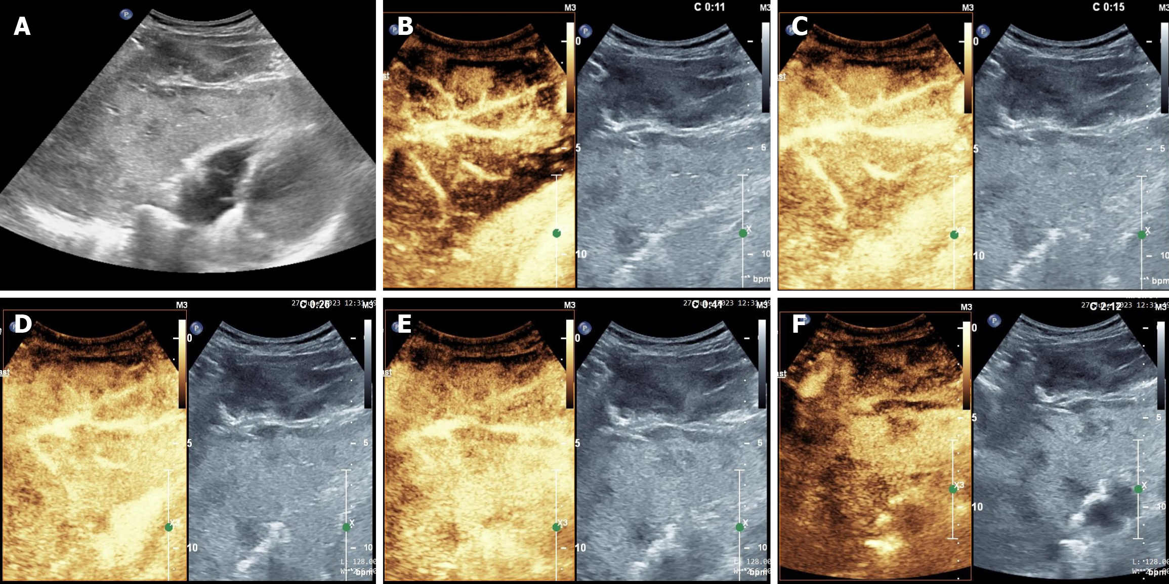
Figure 2 Gray-scale ultrasound and contrast-enhanced ultrasonography of the patient with primary hepatic angiosarcoma.
A: The gray-scale ultrasound image showing a patchy hypoechoic area in the liver with an ill-defined border; B: The manifestation of contrast-enhanced ultrasonography (CEUS) at 11 s of arterial phase; C: The manifestation of CEUS at 15 s of arterial phase; D: The manifestation of CEUS at 26 s of the arterial phase; E: The manifestation of CEUS at 41 s of the portal venous phase; F: The manifestation of CEUS after 120 s of the delayed phase. In the arterial phase, the lamellar hypoechoic area exhibited heterogeneous hyperenhancement along the hepatic pedicle structure, and the contrast agent in this area washed-out mildly and rapidly, with no invasion of the hepatic pedicle structure. In the portal venous phase, the area showed slight hypoenhancement, and well-defined hepatic pedicle structures were visible. In the delayed phase, the enhanced region changed to significant hypoenhancement and was significantly lower than the surrounding normal liver parenchyma.
- Citation: Lin XJ, Luo HC. Rare infiltrative primary hepatic angiosarcoma: A case report and review of literature. World J Gastrointest Oncol 2024; 16(7): 3341-3349
- URL: https://www.wjgnet.com/1948-5204/full/v16/i7/3341.htm
- DOI: https://dx.doi.org/10.4251/wjgo.v16.i7.3341