
















©The Author(s) 2024.
World J Gastrointest Oncol. Jul 15, 2024; 16(7): 2941-2951
Published online Jul 15, 2024. doi: 10.4251/wjgo.v16.i7.2941
Published online Jul 15, 2024. doi: 10.4251/wjgo.v16.i7.2941
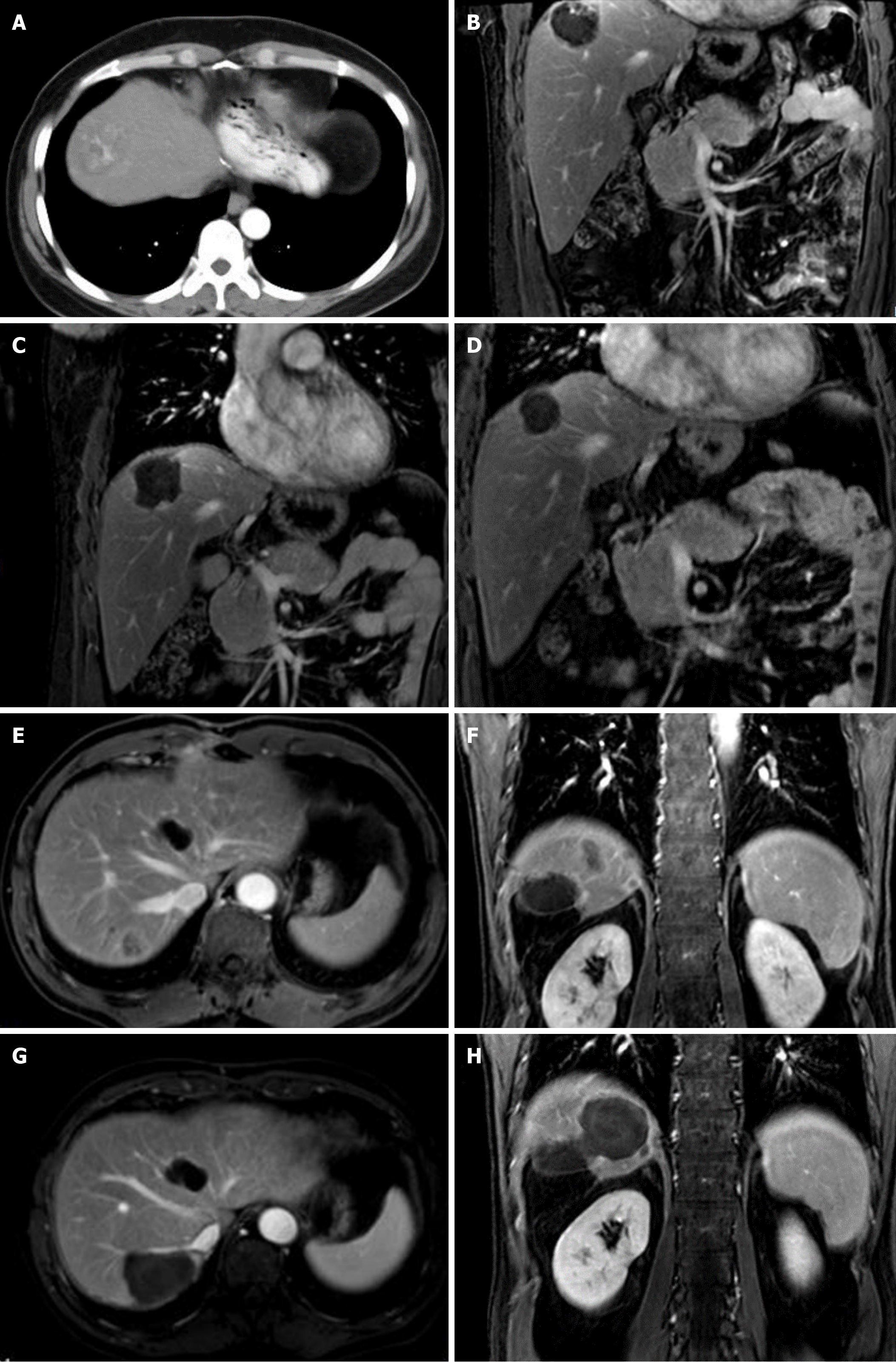
Figure 2 Two patients with cirrhosis and subphrenic hepatocellular carcinoma were treated with transarterial chemoembolization-microwave ablation.
A-D: 50-year-old female with cirrhosis and subphrenic hepatocellular carcinoma measuring 5.0 cm in maximum diameter in segment 8 of the liver. Preprocedure axial contrast computed tomography shows a hypervascular tumor in the liver dome (A); coronal enhanced magnetic resonance imaging (MRI) showing suspicious residues within the upper part of the tumor after transarterial chemotherapy with a combination of 30 mg of epirubicin and 5 mL iodized oil (B); 1 mo after the combination treatment, coronal contrast MRI showed that the ablation area encompassed the tumor and was close to the diaphragm (C); 4 years after the combination treatment, coronal contrast MRI showed no residual tumor and significant shrinkage of the ablation area compared with 1-mo postprocedure (D); E-H: 65-year-male with cirrhosis and subphrenic hepatocellular carcinoma measuring 3.2 cm in maximum diameter in segment 7 of the liver. Preoperative axial and coronal contrast MRI showing a hypervascular tumor in the liver dome (E and F); 1 mo after combination treatment, axial and coronal contrast MRI showed that the ablation area encompassed the tumor and was close to the diaphragm and right hepatic vein (G and H).
- Citation: Zhu ZY, Qian Z, Qin ZQ, Xie B, Wei JZ, Yang PP, Yuan M. Effectiveness and safety of sequential transarterial chemoembolization and microwave ablation for subphrenic hepatocellular carcinoma: A comprehensive evaluation. World J Gastrointest Oncol 2024; 16(7): 2941-2951
- URL: https://www.wjgnet.com/1948-5204/full/v16/i7/2941.htm
- DOI: https://dx.doi.org/10.4251/wjgo.v16.i7.2941