Copyright
©The Author(s) 2024.
World J Gastrointest Oncol. May 15, 2024; 16(5): 2253-2260
Published online May 15, 2024. doi: 10.4251/wjgo.v16.i5.2253
Published online May 15, 2024. doi: 10.4251/wjgo.v16.i5.2253
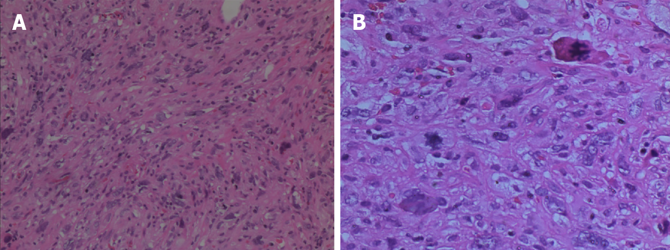
Figure 4 Pathological examination findings.
The tumor cells were spindle-shaped or pleomorphic, arranged in sarciniform structure, with deeply stained nuclei, singular nuclei and nuclear division, abundant cytoplasm, and scattered inflammatory cells infiltration. A: Hematoxylin & eosin (H&E) staining, magnification: 100 ×; B: H&E staining, magnification: 200 ×.
- Citation: Zheng LP, Shen WY, Hu CD, Wang CH, Chen XJ, Wang J, Shen YY. Undifferentiated high-grade pleomorphic sarcoma of the common bile duct: A case report and review of literature. World J Gastrointest Oncol 2024; 16(5): 2253-2260
- URL: https://www.wjgnet.com/1948-5204/full/v16/i5/2253.htm
- DOI: https://dx.doi.org/10.4251/wjgo.v16.i5.2253


