Copyright
©The Author(s) 2024.
World J Gastrointest Oncol. Apr 15, 2024; 16(4): 1647-1659
Published online Apr 15, 2024. doi: 10.4251/wjgo.v16.i4.1647
Published online Apr 15, 2024. doi: 10.4251/wjgo.v16.i4.1647
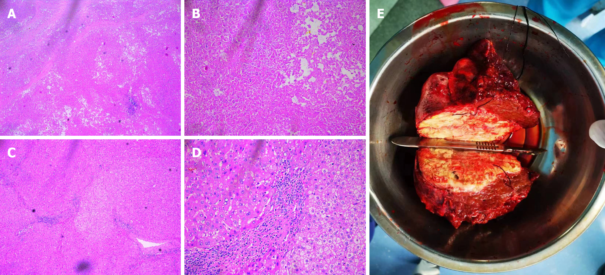
Figure 7 Histopathology of tumor tissue after conversion therapy and resection.
A and B: At low magnification (50 ×) and high magnification (200 ×), the primary lesion showed massive coagulation necrosis with lymphocyte infiltration; C and D: At low magnification (50 ×) and high magnification (200 ×), hyperplasia of the surrounding fibrous tissue and lymphocyte infiltration were visible in nontumor normal tissues; E: Macroscopic view of the patient's intrahepatic lesion.
- Citation: Chu JH, Huang LY, Wang YR, Li J, Han SL, Xi H, Gao WX, Cui YY, Qian MP. Pathologically successful conversion hepatectomy for advanced giant hepatocellular carcinoma after multidisciplinary therapy: A case report and review of literature. World J Gastrointest Oncol 2024; 16(4): 1647-1659
- URL: https://www.wjgnet.com/1948-5204/full/v16/i4/1647.htm
- DOI: https://dx.doi.org/10.4251/wjgo.v16.i4.1647


