
















©The Author(s) 2024.
World J Gastrointest Oncol. Jan 15, 2024; 16(1): 118-132
Published online Jan 15, 2024. doi: 10.4251/wjgo.v16.i1.118
Published online Jan 15, 2024. doi: 10.4251/wjgo.v16.i1.118
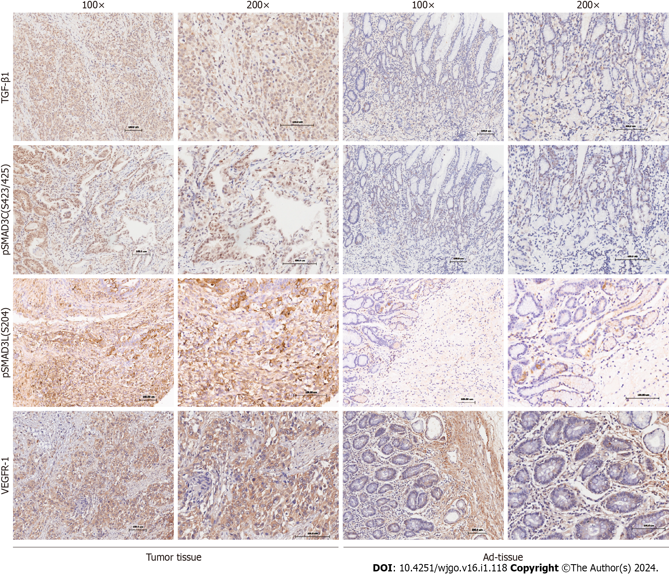
Figure 1 The expression of TGF-β1, pSMAD3C, pSMAD3L, and VEGFR-1 in gastric cancer.
Representative immunohistochemical images showing the expression of TGF-β1, pSMAD3C, pSMAD3L, and VEGFR-1 in cancerous (tumor tissue) and normal (Ad-tissue) gastric tissue. The right panel provides an enlarged view of the specific features highlighted in the rectangular image presented in the left panel. (Scale bar, 100 μm).
- Citation: Lv SL, Guo P, Zou JR, Chen RS, Luo LY, Huang DQ. Prognostic significance and relationship of SMAD3 phospho-isoforms and VEGFR-1 in gastric cancer: A clinicopathological study. World J Gastrointest Oncol 2024; 16(1): 118-132
- URL: https://www.wjgnet.com/1948-5204/full/v16/i1/118.htm
- DOI: https://dx.doi.org/10.4251/wjgo.v16.i1.118