Copyright
©The Author(s) 2023.
World J Gastrointest Oncol. Jan 15, 2023; 15(1): 195-204
Published online Jan 15, 2023. doi: 10.4251/wjgo.v15.i1.195
Published online Jan 15, 2023. doi: 10.4251/wjgo.v15.i1.195
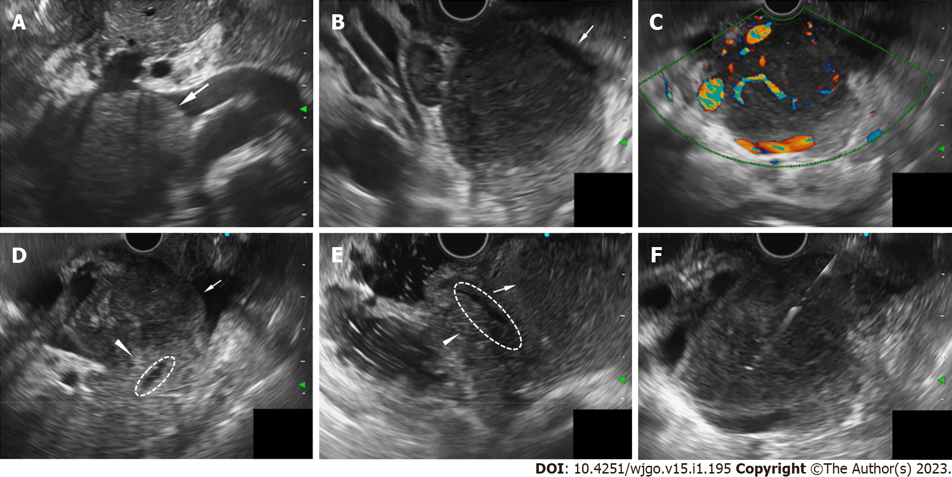
Figure 2 Endoscopic ultrasound characteristic of the lesion located in pancreatic head region.
A and B: A 51.0 mm × 45.8 mm well-defined homogenously hypoechoic lesion located in the pancreatic head region (A: Transgastric observation, white arrow; B: Transduodenal observation, white arrow); C: The lesion is hypervascularity in color and power Doppler ultrasound; D and E: Observing in the duodenal bulb (D) and descending part of duodenum (E), the lesion (white arrow) is adjacent to the pancreatic head parenchyma (white arrow head), but boundaries were still preserved. The main pancreatic duct (dotted circle) ran alongside the lesion naturally, without any signs of infiltration or dilatation; F: Endoscopic ultrasound-Fine needle biopsy using a 22 gauge needle was performed.
- Citation: Wang YN, Zhu YM, Lei XJ, Chen Y, Ni WM, Fu ZW, Pan WS. Intestinal natural killer/T-cell lymphoma presenting as a pancreatic head space-occupying lesion: A case report. World J Gastrointest Oncol 2023; 15(1): 195-204
- URL: https://www.wjgnet.com/1948-5204/full/v15/i1/195.htm
- DOI: https://dx.doi.org/10.4251/wjgo.v15.i1.195


