Copyright
©The Author(s) 2022.
World J Gastrointest Oncol. Aug 15, 2022; 14(8): 1499-1509
Published online Aug 15, 2022. doi: 10.4251/wjgo.v14.i8.1499
Published online Aug 15, 2022. doi: 10.4251/wjgo.v14.i8.1499
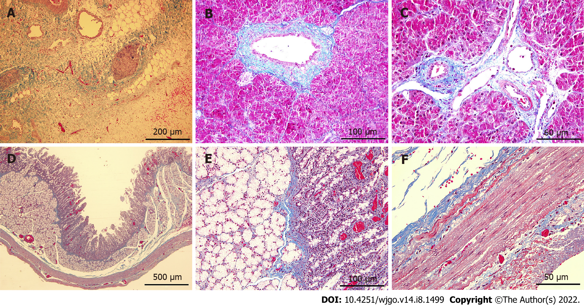
Figure 6 Masson trichrome staining of tissues in the ablation zone.
A-C: Mild fibrosis (blue stained) was observed in pancreatic parenchyma on 7 d post-ablation (A), and the structures of pancreatic ducts (B) and vessels (C) remained intact; D-F: Staining of the duodenum wall showed that the structure of all layers was preserved although minimal injury to the mucosa layer was noted. Scale bars in (A) = 200 μm. Scale bars in (D) = 500 μm. Scale bars in (B and E) = 100 μm. Scale bars in (C and F) = 50 μm.
- Citation: Yan L, Liang B, Feng J, Zhang HY, Chang HS, Liu B, Chen YL. Safety and feasibility of irreversible electroporation for the pancreatic head in a porcine model. World J Gastrointest Oncol 2022; 14(8): 1499-1509
- URL: https://www.wjgnet.com/1948-5204/full/v14/i8/1499.htm
- DOI: https://dx.doi.org/10.4251/wjgo.v14.i8.1499


