Copyright
©The Author(s) 2022.
World J Gastrointest Oncol. Jul 15, 2022; 14(7): 1348-1355
Published online Jul 15, 2022. doi: 10.4251/wjgo.v14.i7.1348
Published online Jul 15, 2022. doi: 10.4251/wjgo.v14.i7.1348
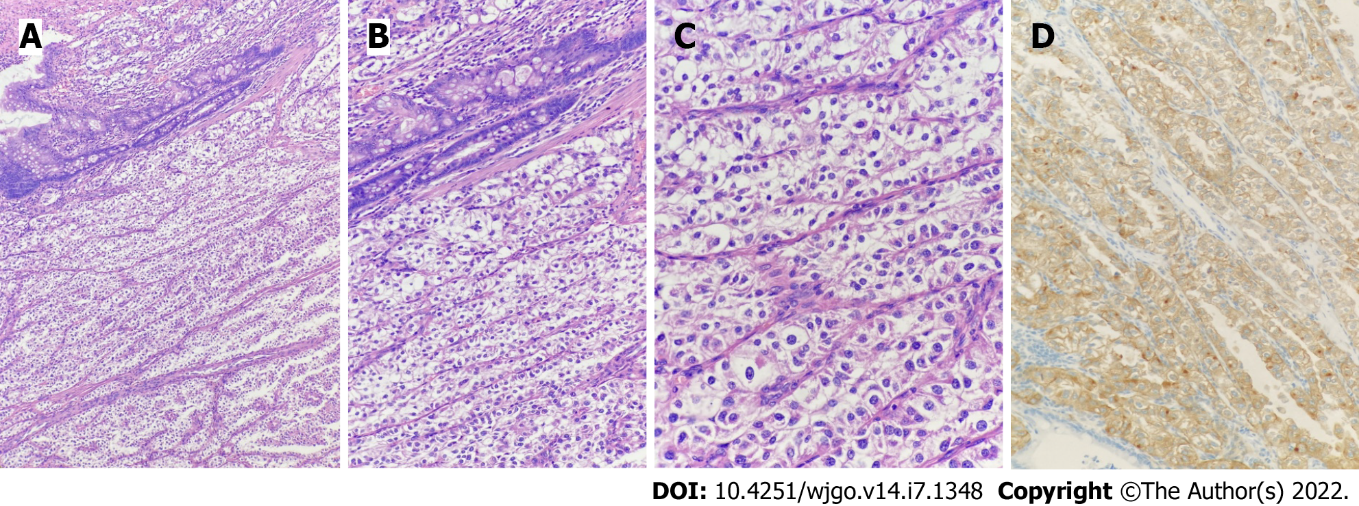
Figure 4 Pathology and immunohistochemistry results.
A: 40 × magnification showing that the tumor was located in the intestinal wall, and the tumor cells were arranged in nests or acini; B: 100 × magnification showing that the tumor cells were transparent or eosinophilic granular; C: 200 × magnification showing abundant capillaries in the interstitium; D: Human melanoma black 45 (+) detected by the EnVision method.
- Citation: Kou L, Zheng WW, Jia L, Wang XL, Zhou JH, Hao JR, Liu Z, Gao FY. Pediatric case of colonic perivascular epithelioid cell tumor complicated with intussusception and anal incarceration: A case report. World J Gastrointest Oncol 2022; 14(7): 1348-1355
- URL: https://www.wjgnet.com/1948-5204/full/v14/i7/1348.htm
- DOI: https://dx.doi.org/10.4251/wjgo.v14.i7.1348


