Copyright
©The Author(s) 2022.
World J Gastrointest Oncol. May 15, 2022; 14(5): 1037-1049
Published online May 15, 2022. doi: 10.4251/wjgo.v14.i5.1037
Published online May 15, 2022. doi: 10.4251/wjgo.v14.i5.1037
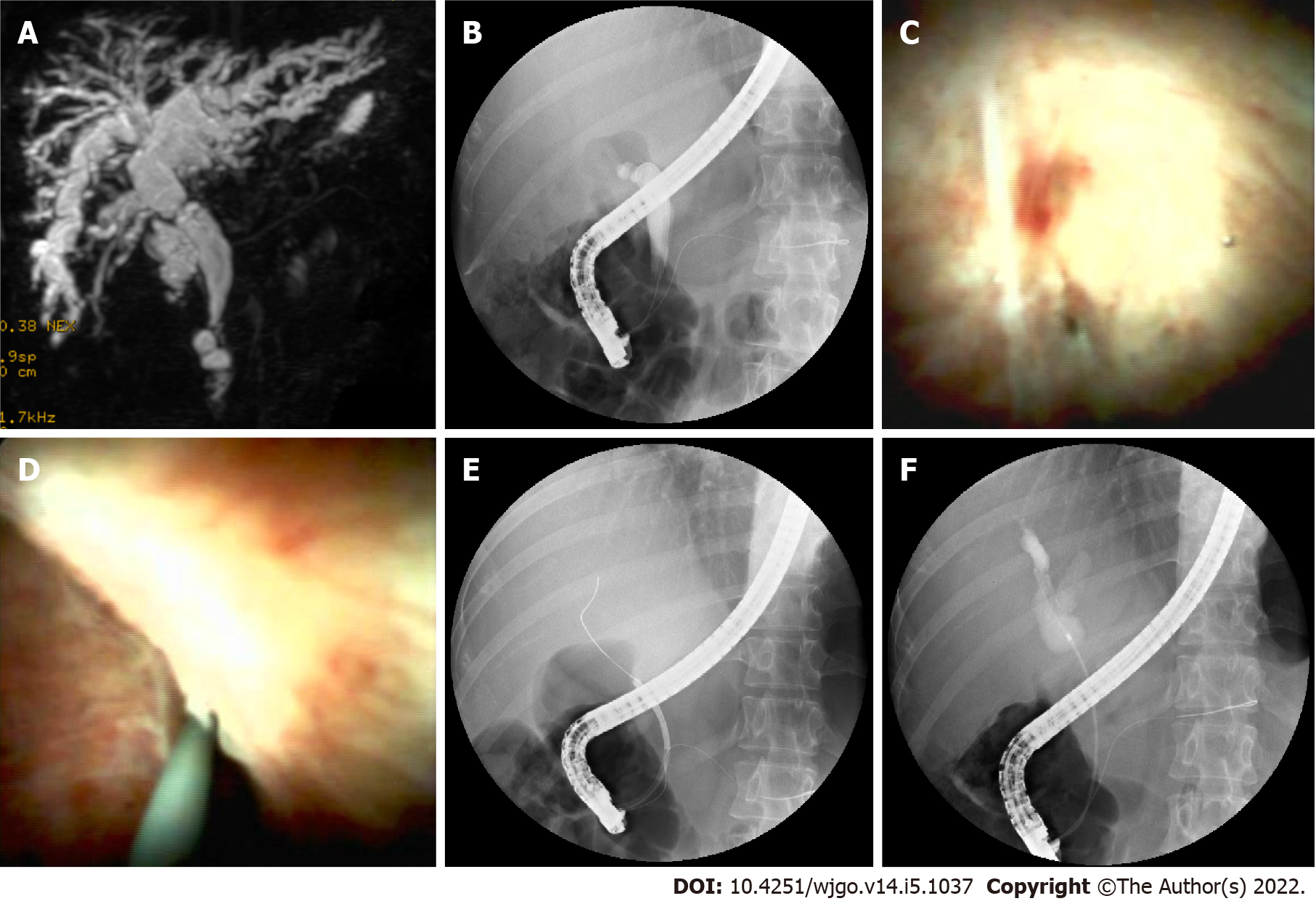
Figure 2 Cholangioscopy-assisted guidewire placement.
A: MRCP image shows anastomotic stricture and dilated bile duct above and below the stricture; B: ERCP image shows the guidewire failed to pass through the stricture; C: A narrow needle-like anastomosis (black arrow); D: Cholangioscopic image shown guidewire inserted through the anastomosis; E: ERCP image shown guidewire inserted into the intrahepatic bile duct; F: ERCP image shown dilated bile duct above anastomosis.
- Citation: Yu JF, Zhang DL, Wang YB, Hao JY. Digital single-operator cholangioscopy for biliary stricture after cadaveric liver transplantation. World J Gastrointest Oncol 2022; 14(5): 1037-1049
- URL: https://www.wjgnet.com/1948-5204/full/v14/i5/1037.htm
- DOI: https://dx.doi.org/10.4251/wjgo.v14.i5.1037


