Copyright
©The Author(s) 2022.
World J Gastrointest Oncol. May 15, 2022; 14(5): 1027-1036
Published online May 15, 2022. doi: 10.4251/wjgo.v14.i5.1027
Published online May 15, 2022. doi: 10.4251/wjgo.v14.i5.1027
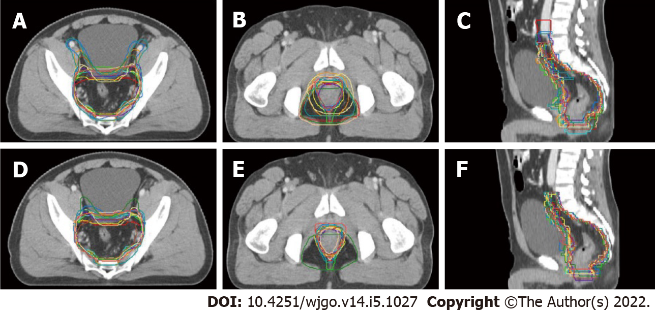
Figure 1 Example images showing the differences in clinical target volume delineation variation before and after the education program.
A: Junction slice of rectum and sigmoid colon (before the education program); B: Slice of the ischiorectal fossa (before the education program); C: Sagittal view (before the education program); D: Junction slice of rectum and sigmoid colon (after the education program); E: Slice of the ischiorectal fossa (after the education program); F: Sagittal view (after the education program). Target volumes delineated by different participants are displayed in different colors.
- Citation: Zhang YZ, Zhu XG, Song MX, Yao KN, Li S, Geng JH, Wang HZ, Li YH, Cai Y, Wang WH. Improving the accuracy and consistency of clinical target volume delineation for rectal cancer by an education program. World J Gastrointest Oncol 2022; 14(5): 1027-1036
- URL: https://www.wjgnet.com/1948-5204/full/v14/i5/1027.htm
- DOI: https://dx.doi.org/10.4251/wjgo.v14.i5.1027


