
















©The Author(s) 2022.
World J Gastrointest Oncol. Mar 15, 2022; 14(3): 587-606
Published online Mar 15, 2022. doi: 10.4251/wjgo.v14.i3.587
Published online Mar 15, 2022. doi: 10.4251/wjgo.v14.i3.587
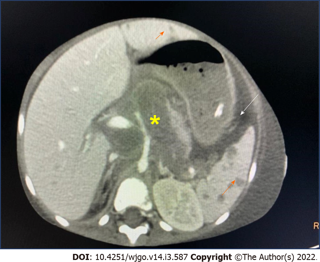
Figure 1 Axial post contrast computed tomography image showing retroperitoneal lymphadenopathy with encasement of celiac artery and portal vein (yellow asterisk).
There are multiple hypoenhancing lesions in liver, spleen (orange arrow) and presence of chylous ascites (white arrow).
- Citation: Devarapalli UV, Sarma MS, Mathiyazhagan G. Gut and liver involvement in pediatric hematolymphoid malignancies. World J Gastrointest Oncol 2022; 14(3): 587-606
- URL: https://www.wjgnet.com/1948-5204/full/v14/i3/587.htm
- DOI: https://dx.doi.org/10.4251/wjgo.v14.i3.587