Copyright
©The Author(s) 2022.
World J Gastrointest Oncol. Dec 15, 2022; 14(12): 2367-2379
Published online Dec 15, 2022. doi: 10.4251/wjgo.v14.i12.2367
Published online Dec 15, 2022. doi: 10.4251/wjgo.v14.i12.2367
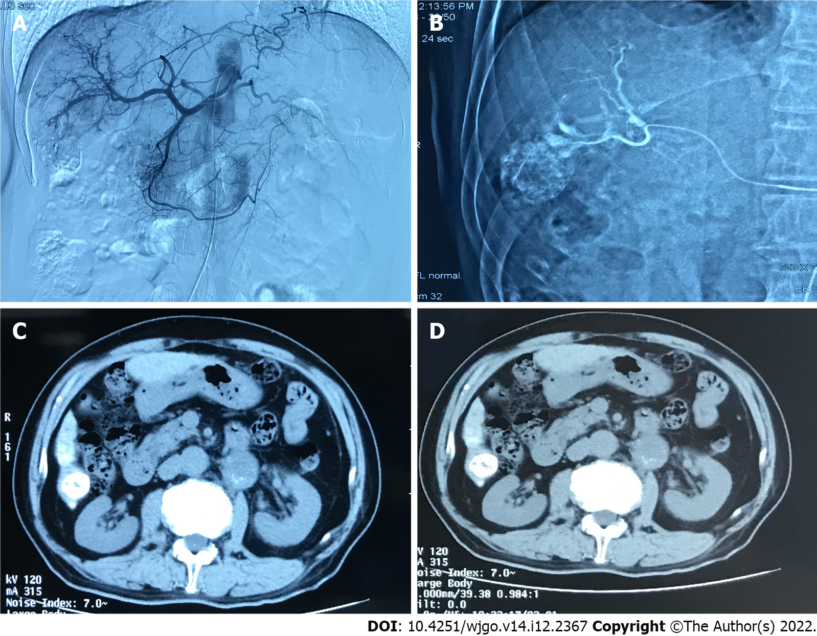
Figure 3 A 70 years old male diagnosed as primary liver cancer.
This patient underwent traditional transcatheter arterial chemoembolization treatment in May 2019. A: Intraoperative digital subtracted angiography (DSA) can be seen on angiography, and the blood supply area of the right hepatic artery can be seen in the lesion; B: Intraoperative DSA, superselective right hepatic artery branch chemoembolization; C: Abdominal computed tomography (CT) axial examination 1 mo after surgery, The lipiodol deposition in the lesion was good, and the lesion was completely necrotic; D: 3 mo after the operation, the abdominal CT axial examination was performed again, and the lipiodol deposition in the lesion was good, and no tumor recurrence was found.
- Citation: Ye T, Shao SH, Ji K, Yao SL. Evaluation of short-term effects of drug-loaded microspheres and traditional transcatheter arterial chemoembolization in the treatment of advanced liver cancer. World J Gastrointest Oncol 2022; 14(12): 2367-2379
- URL: https://www.wjgnet.com/1948-5204/full/v14/i12/2367.htm
- DOI: https://dx.doi.org/10.4251/wjgo.v14.i12.2367


