Copyright
©The Author(s) 2022.
World J Gastrointest Oncol. Dec 15, 2022; 14(12): 2367-2379
Published online Dec 15, 2022. doi: 10.4251/wjgo.v14.i12.2367
Published online Dec 15, 2022. doi: 10.4251/wjgo.v14.i12.2367
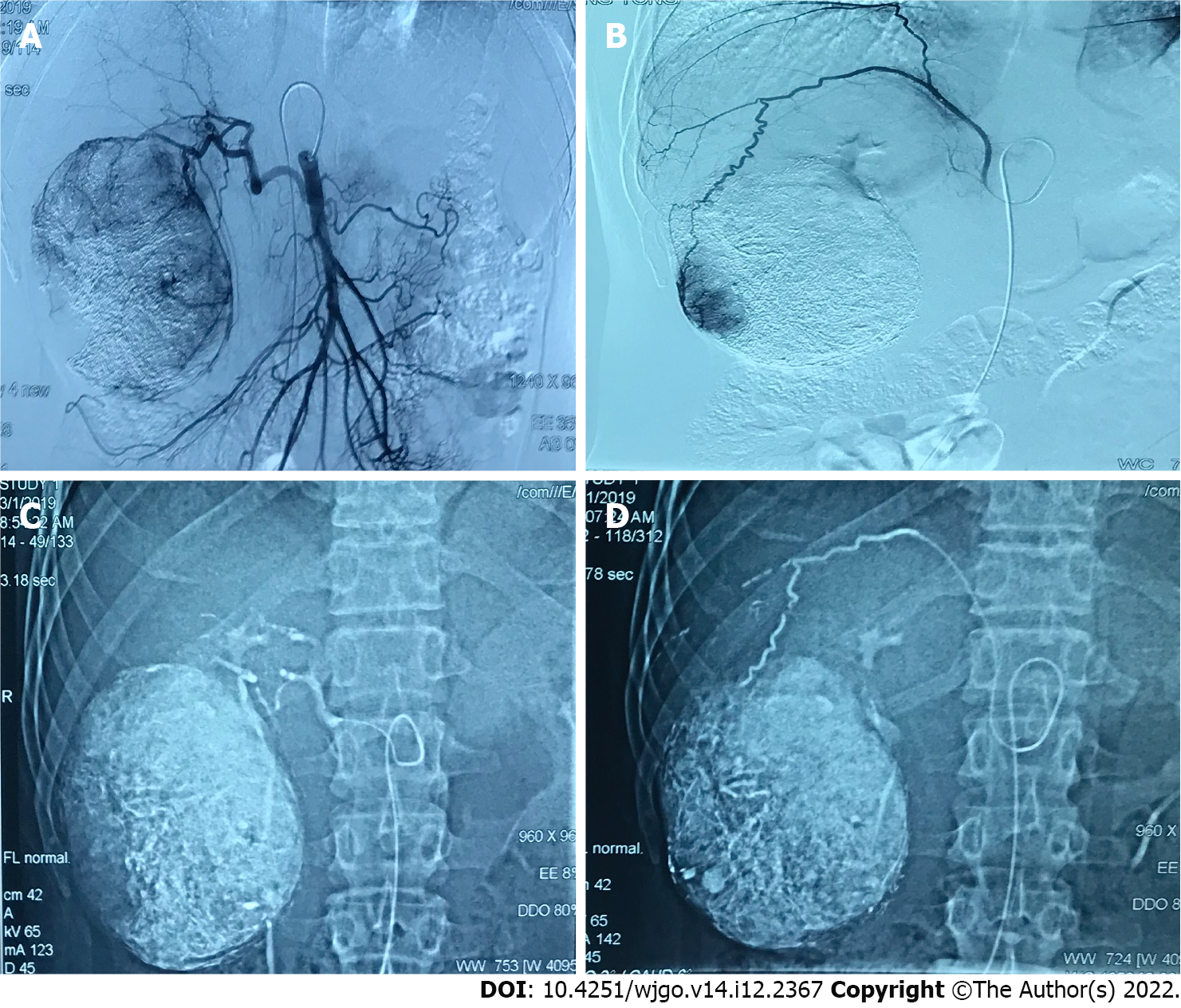
Figure 2 A 38 years old male diagnosed as primary liver cancer.
Traditional transcatheter arterial chemoembolization (TACE) treatment was performed in January 2019, and a follow-up examination was performed one month after the operation. Part of the lesion was alive. Later, the second traditional TACE treatment was performed in March 2019. A: Intraoperative digital subtracted angiography (DSA) angiography shows that the proper hepatic artery originates from the superior mesenteric artery, and the blood supply area of the right hepatic artery can be seen in the lesion; B: Intraoperative DSA angiography shows that the right inferior septal artery participates in the blood supply of the lesion; C: Intraoperative DSA Under the hepatic artery chemoembolization; D: Under DSA during the operation, the right inferior septal artery embolization was performed. Complete necrosis of the lesion was re-examined in January and March postoperative.
- Citation: Ye T, Shao SH, Ji K, Yao SL. Evaluation of short-term effects of drug-loaded microspheres and traditional transcatheter arterial chemoembolization in the treatment of advanced liver cancer. World J Gastrointest Oncol 2022; 14(12): 2367-2379
- URL: https://www.wjgnet.com/1948-5204/full/v14/i12/2367.htm
- DOI: https://dx.doi.org/10.4251/wjgo.v14.i12.2367


