Copyright
©The Author(s) 2021.
World J Gastrointest Oncol. Oct 15, 2021; 13(10): 1302-1316
Published online Oct 15, 2021. doi: 10.4251/wjgo.v13.i10.1302
Published online Oct 15, 2021. doi: 10.4251/wjgo.v13.i10.1302
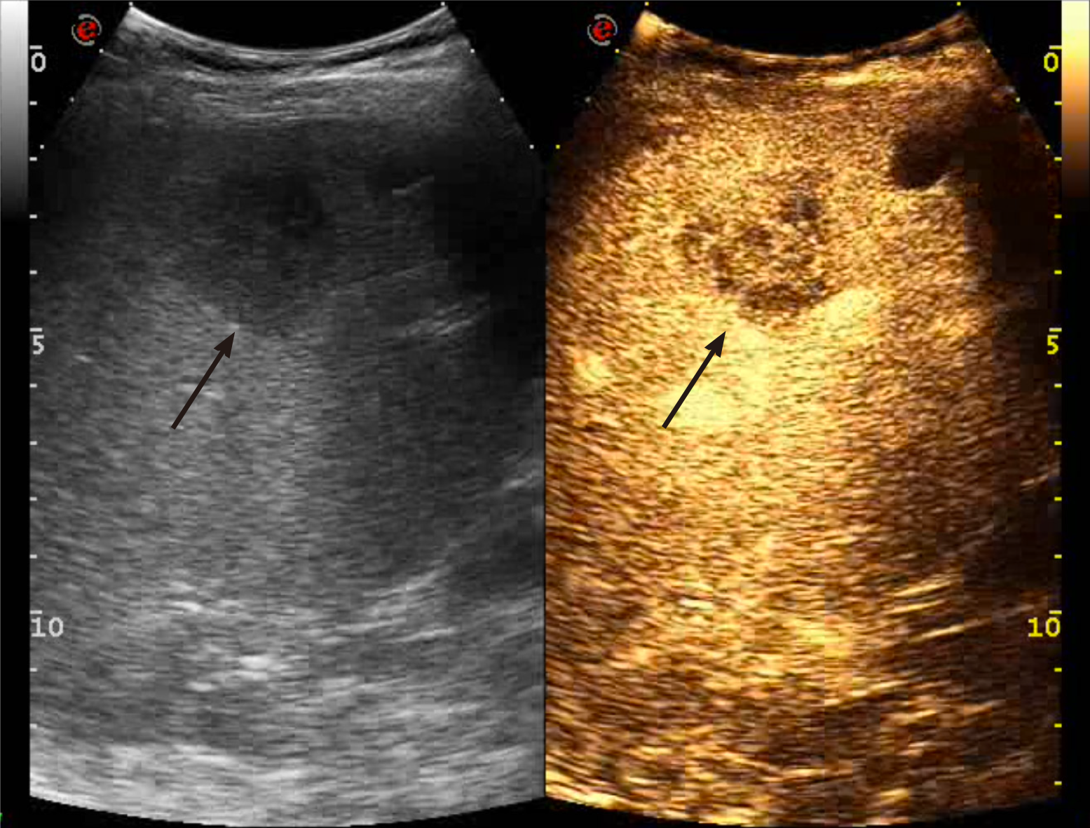
Figure 4 Liver metastasis.
Contrast enhanced ultrasound examination in the early portal phase (50 s after the i.v. injection of contrast agent) shows a heterogeneously vascularized mass, hypoechoic to the surrounding liver parenchyma (black arrow). An anechoic simple cyst is located nearby (black arrow).
- Citation: Bartolotta TV, Taibbi A, Randazzo A, Gagliardo C. New frontiers in liver ultrasound: From mono to multi parametricity. World J Gastrointest Oncol 2021; 13(10): 1302-1316
- URL: https://www.wjgnet.com/1948-5204/full/v13/i10/1302.htm
- DOI: https://dx.doi.org/10.4251/wjgo.v13.i10.1302


