Copyright
©The Author(s) 2020.
World J Gastrointest Oncol. Aug 15, 2020; 12(8): 893-902
Published online Aug 15, 2020. doi: 10.4251/wjgo.v12.i8.893
Published online Aug 15, 2020. doi: 10.4251/wjgo.v12.i8.893
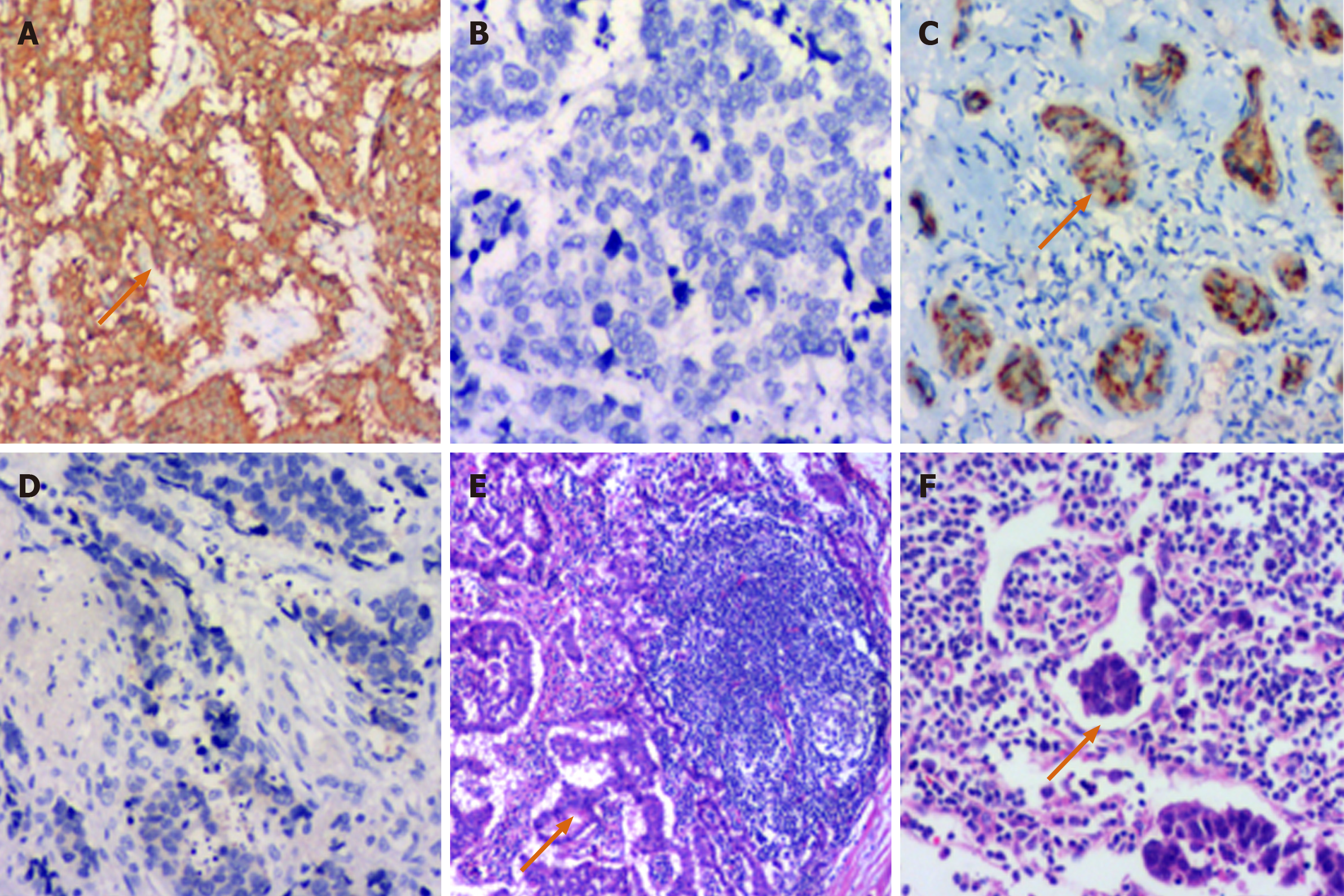
Figure 2 Immunohistochemical features.
A: Positive synaptophysin immunohistochemical staining in a G2 rectal neuroendocrine tumor (RNET) (black arrow heads the positive staining; × 100). B: Negative synaptophysin immunohistochemical staining in an RNEC (× 100); C: Positive chromogranin A immunohistochemical staining in a G1 RNET (black arrow heads the positive staining; × 100); D: Negative chromogranin A immunohistochemical staining in an RNEC (× 100); E: Lymph node metastasis of a G2 RNET (black arrow shows the tumor tissue; hematoxylin and eosin staining, × 100); F: Black arrow shows a thrombus in the lymph vessel (hematoxylin and eosin staining, × 100).
- Citation: Yu YJ, Li YW, Shi Y, Zhang Z, Zheng MY, Zhang SW. Clinical and pathological characteristics and prognosis of 132 cases of rectal neuroendocrine tumors. World J Gastrointest Oncol 2020; 12(8): 893-902
- URL: https://www.wjgnet.com/1948-5204/full/v12/i8/893.htm
- DOI: https://dx.doi.org/10.4251/wjgo.v12.i8.893


