Copyright
©The Author(s) 2020.
World J Gastrointest Oncol. Jul 15, 2020; 12(7): 768-781
Published online Jul 15, 2020. doi: 10.4251/wjgo.v12.i7.768
Published online Jul 15, 2020. doi: 10.4251/wjgo.v12.i7.768
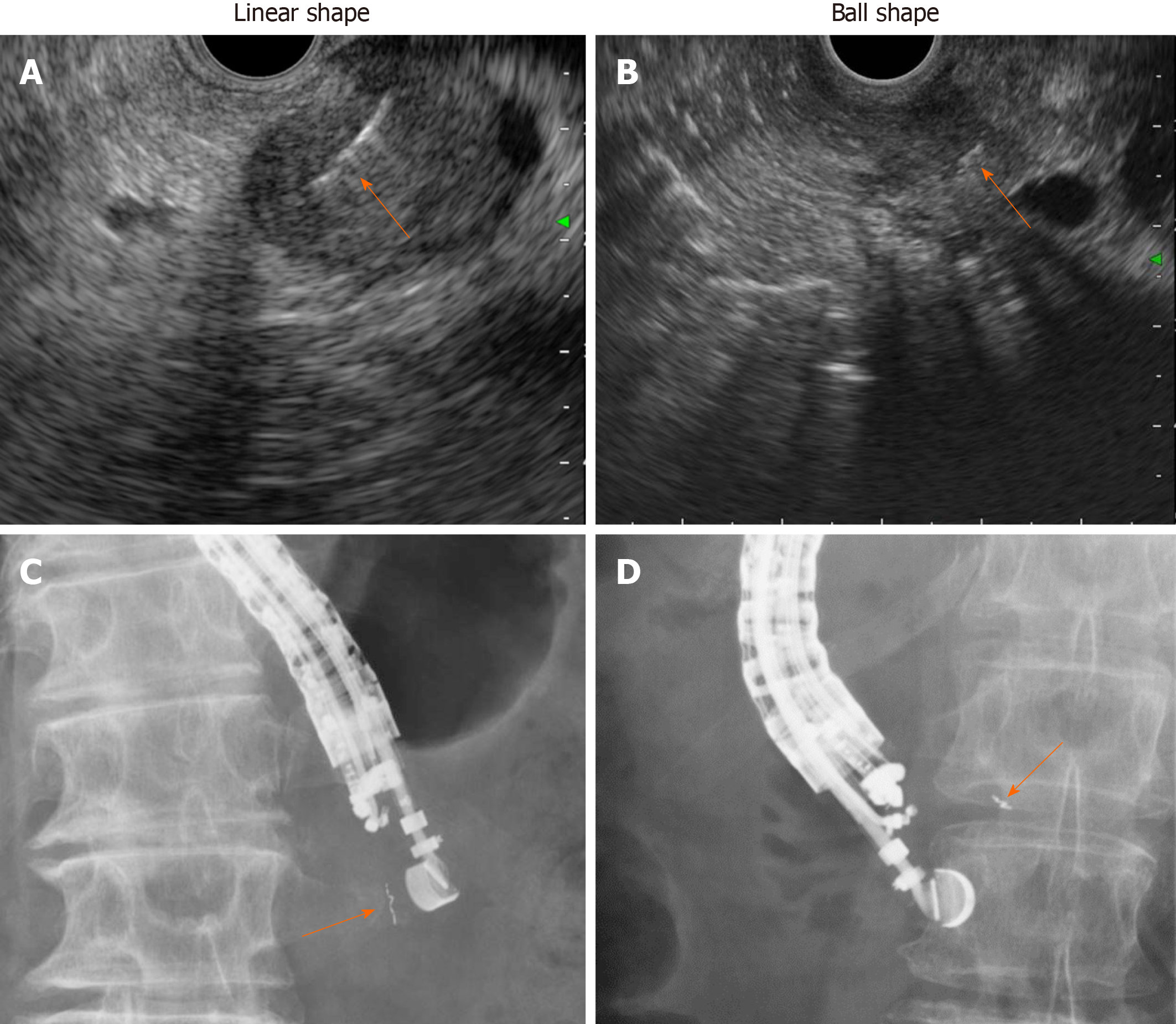
Figure 7 Various insertion techniques for gold anchor.
Linear shape insertion. A: Endoscopic ultrasound image for linear shape insertion; B: Endoscopic ultrasound image for ball shape insertion; C: X-ray image for linear shape insertion. D: X-ray image for ball shape insertion. Orange arrow indicates gold anchor placed in either linear (A, C) or ball (B, D) shape.
- Citation: Ashida R, Fukutake N, Takada R, Ioka T, Ohkawa K, Katayama K, Akita H, Takahashi H, Ohira S, Teshima T. Endoscopic ultrasound-guided fiducial marker placement for neoadjuvant chemoradiation therapy for resectable pancreatic cancer. World J Gastrointest Oncol 2020; 12(7): 768-781
- URL: https://www.wjgnet.com/1948-5204/full/v12/i7/768.htm
- DOI: https://dx.doi.org/10.4251/wjgo.v12.i7.768


